. 2009 Jun;77(6):2508-16.
doi: 10.1128/IAI.00166-09. Epub 2009 Mar 23.

Cytolethal distending toxin promotes Helicobacter cinaedi-associated typhlocolitis in interleukin-10-deficient mice

Affiliations

Cytolethal distending toxin promotes Helicobacter cinaedi-associated typhlocolitis in interleukin-10-deficient mice

Z Shen et al. Infect Immun. 2009 Jun.

Abstract

Helicobacter cinaedi colonizes a wide host range, including rodents, and may be an emerging zoonotic agent. Colonization parameters, pathology, serology, and inflammatory responses to wild-type H. cinaedi (WT(Hc)) were evaluated in B6.129P2-IL-10(tm1Cgn) (IL-10(-/-)) mice for 36 weeks postinfection (WPI) and in C57BL/6 (B6) mice for 12 WPI. Because cytolethal distending toxin (CDT) may be a virulence factor, IL-10(-/-) mice were also infected with the cdtB(Hc) and cdtB-N(Hc) isogenic mutants and evaluated for 12 WPI. Consistent with other murine enterohepatic helicobacters, WT(Hc) did not cause typhlocolitis in B6 mice, but mild to severe lesions developed at the cecocolic junction in IL-10(-/-) mice, despite similar colonization levels of WT(Hc) in the cecum and colon of both B6 and IL-10(-/-) mice. WT(Hc) and cdtB mutants also colonized IL-10(-/-) mice to a similar extent, but infection with either cdtB mutant resulted in attenuated typhlocolitis and hyperplasia compared to infection with WT(Hc) (P < 0.03), and only WT(Hc) infection caused dysplasia and intramucosal carcinoma. WT(Hc) and cdtB(Hc) mutant infection of IL-10(-/-) mice elevated mRNA expression of tumor necrosis factor alpha, inducible nitric oxide synthase, and gamma interferon in the cecum, as well as elevated Th1-associated serum immunoglobulin G2a(b) compared to infection of B6 mice (P < 0.05). Although no hepatitis was noted, liver samples were PCR positive at various time points for WT(Hc) or the cdtB(Hc) mutant in approximately 33% of IL-10(-/-) mice and in 10 to 20% of WT(Hc)-infected B6 mice. These results indicate that WT(Hc) can be used to model inflammatory bowel disease in IL-10(-/-) mice and that CDT contributes to the virulence of H. cinaedi.


Figures

FIG. 1.
FIG. 1.
Giemsa-stained HeLa S3 cells exposed for 72 h to filter-sterilized supernatant of cell sonicates of the H. cinaedi WT strain and cdtBHc mutant. (Magnification, ×200.) (A) PBS control. (B) WTHc expressed CDT, which induced the formation of giant mono- and multinucleated cells with large nuclei. (C) The isogenic mutant (cdtBHc) lost CDT activity. Treated cells maintained a similar morphology to that of the PBS-treated control cells. Loss of activity was also observed using the second isogenic mutant, cdtB-NHc (not shown).
FIG. 2.
FIG. 2.
H. cinaedi colonization levels of WTHc or the cdtBHc mutant in the cecum of infected mice measured by quantitative PCR analysis at 6, 12, 24, and 36 WPI. Numbers represent the ng of H. cinaedi DNA per μg mouse DNA. Only WTHc and the sham control were evaluated at 24 and 36 WPI. *, P < 0.05. Bars, standard errors.
FIG. 3.
FIG. 3.
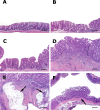
Histopathology of H. cinaedi typhlocolitis in IL-10−/− mice. (A) Cecum of sham-inoculated, disease-free control. (B) Mild or moderate inflammation in a mouse infected with the cdtBHc mutant. (C) Moderate typhlocolitis with mild hyperplasia, dysplasia, and crypt atrophy in a mouse infected with WTHc. (D and E) WTHc-induced gastrointestinal intraepithelial neoplasia with intramuscular dysplastic glands and deep invasion of dilated mucinous glands (arrows). (F) Focal area of epithelial hyperplasia and dysplasia bounded by normal mucosa (skip lesion) in mid-colon of a mouse infected with WTHc. Tissues were stained with hematoxylin and eosin. Bar = 200 μm (A to D and F) and 400 μm (E).
FIG. 4.
FIG. 4.
WTHc or cdtBHc mutant-induced pathology in IL-10−/− mice at 6, 12, 24, and 36 WPI. The median score (horizontal bar) is indicated for each group. Only WTHc and the sham control were evaluated at 24 and 36 WPI. (A) Inflammation. (B) Hyperplasia. (C) Dysplasia. All infected mice had significantly higher inflammation scores than control mice (P < 0.001) at all time points. At 12 WPI, the cdtBHc mutant induced less inflammation than WTHc (*, P < 0.05).
FIG. 5.
FIG. 5.
Median pathology scores of the third experiment using a second isogenic mutant (cdtB-NHc) or WTHc in IL-10−/− mice at 12 WPI. All infected mice had significantly higher inflammation scores than control mice (P < 0.001), and the cdtB-NHc mutant induced less pathology than WTHc (*, P < 0.05). Horizontal bars, median scores.
FIG. 6.
FIG. 6.
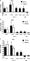
Relative expression of select cytokine and iNOS mRNA levels in the cecum. Expression levels of TNF-α, IFN-γ, and iNOS were higher in infected IL-10−/− mice than those in B6 mice at 12 WPI. Data represent means and standard errors (bars) of changes in mRNA expression in infected mice compared to expression in uninfected controls. *, P < 0.05; **, P < 0.01, compared with the sham control. Only WTHc and the sham control were evaluated at 24 and 36 WPI. There were no statistically significant differences in cytokine levels between WTHc- and the cdtBHc mutant-infected mice.
FIG. 7.
FIG. 7.
Scanning laser confocal immunofluorescent detection of H. cinaedi-induced iNOS and macrophage F4/80 expression in cecal sections of WTHc-infected and uninfected mice. iNOS was upregulated in focal areas in the cecum of WTHc-infected IL-10−/− mice. Green, F4/80; red, iNOS; blue, nucleus. (A) Control mouse. (B and C) WTHc-infected mice.
FIG. 8.
FIG. 8.
Serum antibody levels in infected IL-10 −/− and B6 mice against H. cinaedi whole-cell sonicate protein measured by ELISA at 6 and 12 WPI. (A) IgG1 antibody response. (B) IgG2ab antibody response. Infection with WTHc or the cdtBHc mutant promoted significant IgG2ab (Th1-associated) and IgG1 (Th2-associated) antibody responses by 6 WPI and increased between 6 and 12 WPI. IgG2ab response was higher in infected IL-10−/− mice than in infected B6 mice. *, P < 0.05. Bars, standard errors.

Similar articles

Cited by

References

    1. Boivin, G. P., K. Washington, and K. Yang. 2003. Pathology of mouse models of intestinal cancer: consensus report and recommendations. Gastoenterology 124762-777. - PubMed
    1. Burman, W. J., D. L. Cohn, R. R. Reves, and M. L. Wilson. 1995. Multifocal cellulitis and monoarticular arthritis as manifestations of Helicobacter cinaedi bacteremia. Clin. Infect. Dis. 20564-570. - PubMed
    1. Cahill, R. J., C. J. Foltz, J. G. Fox, C. A. Dangler, F. Powrie, and D. B. Schauer. 1997. Inflammatory bowel disease: an immunity-mediated condition triggered by bacterial infection with Helicobacter hepaticus. Infect. Immun. 653126-3131. - PMC - PubMed
    1. Chien, C. C., N. S. Taylor, Z. Ge, D. B. Schauer, V. B. Young, and J. G. Fox. 2000. Identification of cdtB homologues and cytolethal distending toxin activity in enterohepatic Helicobacter spp. J. Med. Microbiol. 49525-534. - PubMed
    1. Comayras, C., C. Tasca, S. Y. Peres, B. Ducommun, E. Oswald, and J. De Rycke. 1997. Escherichia coli cytolethal distending toxin blocks the HeLa cell cycle at the G2/M transition by preventing cdc2 protein kinase dephosphorylation and activation. Infect. Immun. 655088-5095. - PMC - PubMed

Publication types