. 2009 Jun;77(6):2262-71.
doi: 10.1128/IAI.01324-08. Epub 2009 Mar 23.

Resistance of Capnocytophaga canimorsus to killing by human complement and polymorphonuclear leukocytes

Affiliations

Resistance of Capnocytophaga canimorsus to killing by human complement and polymorphonuclear leukocytes

Hwain Shin et al. Infect Immun. 2009 Jun.

Abstract

Capnocytophaga canimorsus is a bacterium of the canine oral flora known since 1976 to cause rare but severe septicemia and peripheral gangrene in patients that have been in contact with a dog. It was recently shown that these bacteria do not elicit an inflammatory response (H. Shin, M. Mally, M. Kuhn, C. Paroz, and G. R. Cornelis, J. Infect. Dis. 195:375-386, 2007). Here, we analyze their sensitivity to the innate immune system. Bacteria from the archetype strain Cc5 were highly resistant to killing by complement. There was little membrane attack complex (MAC) deposition in spite of C3b deposition. Cc5 bacteria were as resistant to phagocytosis by human polymorphonuclear leukocytes (PMNs) as Yersinia enterocolitica MRS40, endowed with an antiphagocytic type III secretion system. We isolated Y1C12, a transposon mutant that is hypersensitive to killing by complement via the antibody-dependent classical pathway. The mutation inactivated a putative glycosyltransferase gene, suggesting that the Y1C12 mutant was affected at the level of a capsular polysaccharide or lipopolysaccharide (LPS) structure. Cc5 appeared to have several polysaccharidic structures, one being altered in Y1C12. The structure missing in Y1C12 could be purified by classical LPS purification procedures and labeled by tritiated palmitate, indicating that it is more likely to be an LPS structure than a capsule. Y1C12 bacteria were also more sensitive to phagocytosis by PMNs than wild-type bacteria. In conclusion, a polysaccharide structure, likely an LPS, protects C. canimorsus from deposition of the complement MAC and from efficient phagocytosis by PMNs.


Figures

FIG. 1.
FIG. 1.
C. canimorsus cells are resistant to complement-mediated killing. (A) Total CFU present after incubation of Cc5 (black circles), E. coli (white circles), and wt S. enterica serovar Typhimurium (gray circles) in 10% NHS for 0, 15, 30, 60, 120, and 180 min at 37°C. (B) Survival of Cc11 (black circles) and Cc12 (white circles) in 10% NHS for 0, 15, 30, 60, 120, and 180 min at 37°C. (C) Total CFU present after incubation of Cc5 in 10% HI (black) or active (white) normal mouse serum (NMS) or normal dog serum (NDS) for 180 min at 37°C. (D) Binding of C3b by Cc5 or E. coli after 30 min incubation with HI NHS. FACS analysis was performed using anti-C3c polyclonal antiserum to quantify the level of C3b on the bacterial surface. The values are the means of three independent experiments ± standard deviations (bars).
FIG. 2.
FIG. 2.
Cc5 cells are resistant to PMN-mediated killing. Levels of phagocytosis and killing were determined 120 min after infection of human PMNs. PMNs were infected with either Cc5 or wt Y. enterocolitica at an MOI of 1. Infection with nonopsonized (black), HI NHS-opsonized (gray), or C7-depleted NHS-opsonized bacteria (white) are shown. Results are the means of at least three independent experiments ± standard deviations (bars). The statistical significance of the difference between Cc5 and wt Y. enterocolitica (unopsonized) is given, with * indicating a P value of <0.05 and ** indicating a P value of <0.01, using a two-tailed unpaired Student's t test.
FIG. 3.
FIG. 3.
Characterization of the serum-sensitive Tn mutant of Cc5. (A) Total CFU present after incubation of Cc5 (black circles), Y1C12 (white circles), or an S. enterica serovar Typhimurium rfaG mutant (gray circles) in 10% NHS for 0, 15, 30, 60, 120, and 180 min at 37°C. (B) Total CFU present after incubation of Cc5, Y1C12, cY1C12, or an S. enterica serovar Typhimurium rfaG mutant in 10% HI NHS (black) or 10% NHS (white) for 180 min at 37°C. (C) Binding of C3b by Cc5, Y1C12, or cY1C12 after 30 min incubation with C7-depleted NHS. FACS analysis was performed using anti-C3c polyclonal antiserum to quantify the level of C3b on the bacterial surface. (D) MAC deposition on Cc5, Y1C12, or cY1C12 after 15 min of incubation with NHS (longer incubation leads to significant lysis). FACS analysis was performed using mouse anti-C5B-9 primary and anti-mouse-FITC secondary Abs to quantify the level of MAC insertion into the bacterial surface. The statistical significance of the difference in MAC insertion between Y1C12 and wt C. canimorsus is given, with * indicating a P value of <0.05 using a two-tailed unpaired Student's t test. Results are the means of at least three independent experiments ± standard deviations (bars). cY1C12, Y1C12 mutant bacteria complemented with the gtf gene in trans.
FIG. 4.
FIG. 4.
Killing of Y1C12 occurs via an IgG-dependent classical pathway. (A) Total CFU present after incubation of wt or Y1C12 C. canimorsus in 10% HI NHS (black), NHS (dark gray), Ab-depleted NHS (light gray), or Ab-depleted NHS supplemented with HI NHS as a source of Abs (white) for 180 min at 37°C. (B) Total CFU present after incubation of wt or Y1C12 C. canimorsus in 10% HI NHS (black), NHS (gray), or Mg-EGTA-treated NHS (white) for 180 min at 37°C. (C) Binding of human serum Abs to wt, Y1C12, or cY1C12 C. canimorsus after 30 min incubation with HI NHS. The level of Ab deposition on bacteria was quantified by FACS analysis using anti-human IgG-FITC Ab. Results are the means of at least three independent experiments ± standard deviations (bars).
FIG. 5.
FIG. 5.
Tn mutant Y1C12 cells exhibit increased sensitivity to phagocytosis and killing by PMNs. Phagocytosis and killing were determined 120 min after infection of human PMNs. PMNs were infected with wt, Y1C12, or cY1C12 C. canimorsus at an MOI of 1. Infections with nonopsonized (black), HI NHS-opsonized (gray), or C7-depleted NHS-opsonized bacteria (white) are shown. Results are the means of at least three independent experiments ± standard deviations (bars), including statistical significance between phagocytosis and killing of the wt and Y1C12 (unopsonized), with *** indicating a P value of <0.001 using a two-tailed, unpaired Student's t test.
FIG. 6.
FIG. 6.
Y1C12 is affected in a gene encoding a putative glycosyltransferase. Shown is the genetic locus of Y1C12 including its upstream and downstream genes (GenBank accession number FJ214098).
FIG. 7.
FIG. 7.
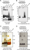
Y1C12 has an altered lipidated polysaccharide structure. (A) Immunoblotting analysis of proteinase K-treated Cc5, Y1C12, and cY1C12 bacteria and of LPS isolated from Cc5 and Y1C12 using anti-Cc5. (B) Immunoblotting analysis as described above (A) using Y1C12-absorbed anti-Cc5 antiserum. (C) Silver-periodic acid staining of Tricine gel electrophoresis of the same samples as above (A and B). (D) Proteinase K-resistant structures of wt Cc5, Y1C12, and cY1C12 bacteria that had been labeled in vivo using [9,10-3H]palmitic acid.

Similar articles

Cited by

References

    1. Bailie, W. E., E. C. Stowe, and A. M. Schmitt. 1978. Aerobic bacterial flora of oral and nasal fluids of canines with reference to bacteria associated with bites. J. Clin. Microbiol. 7223-231. - PMC - PubMed
    1. Blanche, P., E. Bloch, and D. Sicard. 1998. Capnocytophaga canimorsus in the oral flora of dogs and cats. J. Infect. 36134. - PubMed
    1. Bobo, R. A., and E. J. Newton. 1976. A previously undescribed gram-negative bacillus causing septicemia and meningitis. Am. J. Clin. Pathol. 65564-569. - PubMed
    1. Brenner, D. J., D. G. Hollis, G. R. Fanning, and R. E. Weaver. 1989. Capnocytophaga canimorsus sp. nov. (formerly CDC group DF-2), a cause of septicemia following dog bite, and C. cynodegmi sp. nov., a cause of localized wound infection following dog bite. J. Clin. Microbiol. 27231-235. - PMC - PubMed
    1. Cornelis, G., J. C. Vanootegem, and C. Sluiters. 1987. Transcription of the yop regulon from Y. enterocolitica requires trans acting pYV and chromosomal genes. Microb. Pathog. 2367-379. - PubMed

Publication types

Associated data

LinkOut - more resources