. 2009 May;29(10):2762-76.
doi: 10.1128/MCB.01393-08. Epub 2009 Mar 23.

The RNA-binding protein Elavl1/HuR is essential for placental branching morphogenesis and embryonic development

Affiliations

The RNA-binding protein Elavl1/HuR is essential for placental branching morphogenesis and embryonic development

Vicky Katsanou et al. Mol Cell Biol. 2009 May.

Abstract

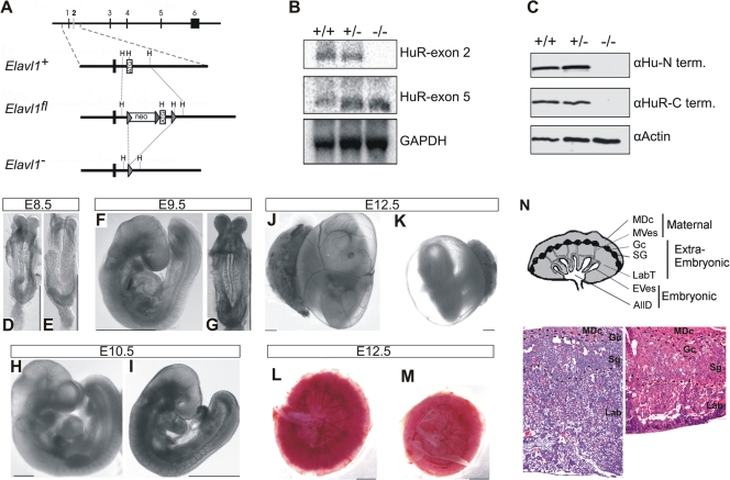
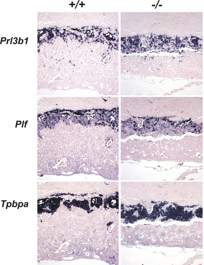
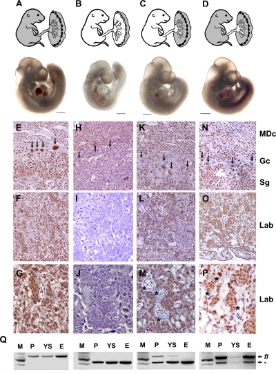
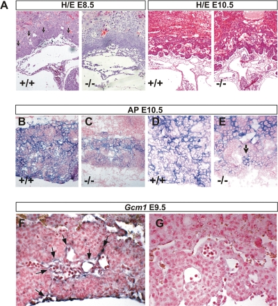
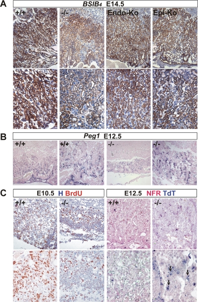
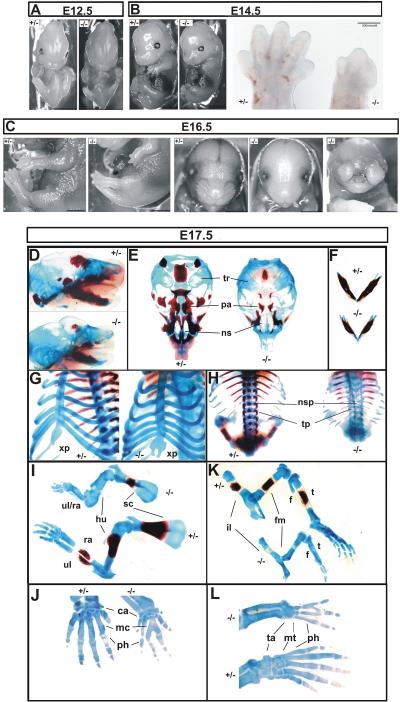
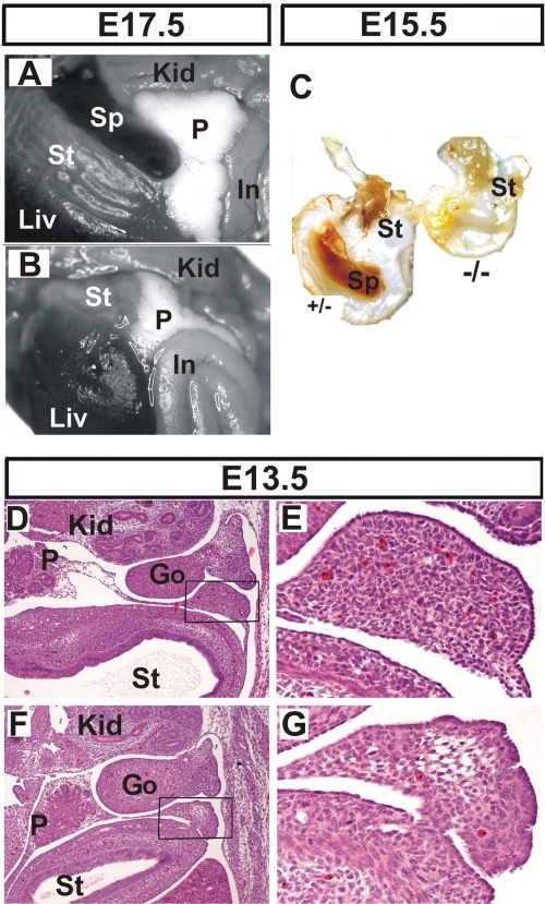
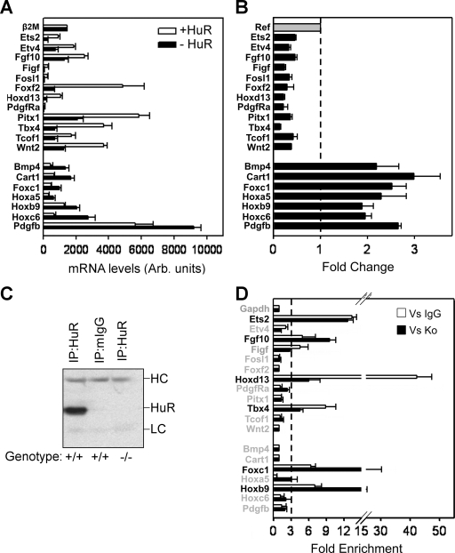
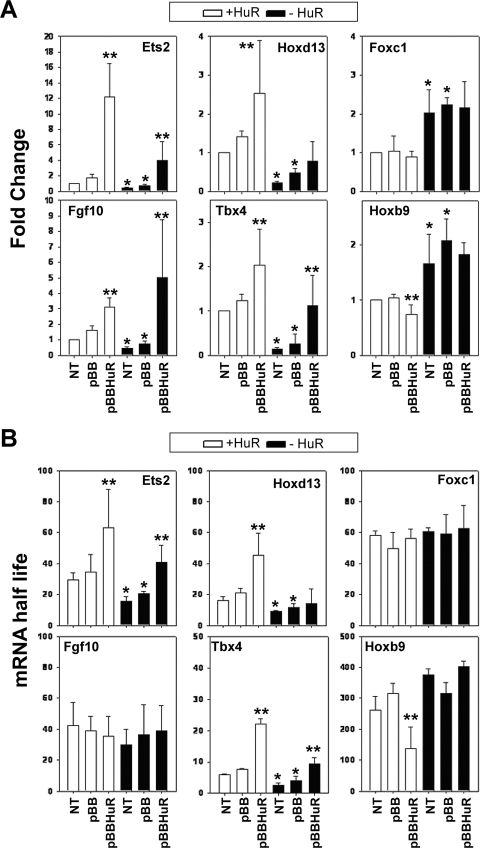
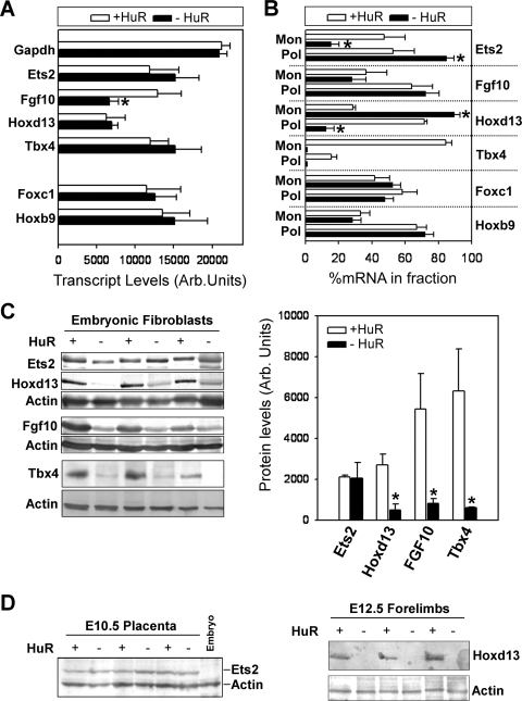
HuR is an RNA-binding protein implicated in a diverse array of pathophysiological processes due to its effects on the posttranscriptional regulation of AU- and U-rich mRNAs. Here we reveal HuR's requirement in embryonic development through its genetic ablation. Obligatory HuR-null embryos exhibited a stage retardation phenotype and failed to survive beyond midgestation. By means of conditional transgenesis, we restricted HuR's mutation in either embryonic or endothelial compartments to demonstrate that embryonic lethality is consequent to defects in extraembryonic placenta. HuR's absence impaired the invagination of allantoic capillaries into the chorionic trophoblast layer and the differentiation of syncytiotrophoblast cells that control the morphogenesis and vascularization of the placental labyrinth and fetal support. HuR-null embryos rescued from these placental defects proceeded to subsequent developmental stages but displayed defects in skeletal ossification, fusions in limb elements, and asplenia. By coupling gene expression measurements, data meta-analysis, and HuR-RNA association assays, we identified transcription and growth factor mRNAs controlled by HuR, primarily at the posttranscriptional level, to guide morphogenesis, specification, and patterning. Collectively, our data demonstrate the dominant role of HuR in organizing gene expression programs guiding placental labyrinth morphogenesis, skeletal specification patterns, and splenic ontogeny.


Figures

FIG. 1.
FIG. 1.
Midgestational phenotype of HuR-deficient embryos. (A) Schematic of the complete exon/intron orientation of the Elavl1 locus on mouse chromosome 8 and magnifications of the region containing the second exon (white box) as a wild-type (Elavl1+), targeted (Elavl1fl), and inactive, Cre-recombined locus (Elavl1). The neo selection gene and loxP genes (arrowheads) are indicated. Restriction sites for HindIII (H) are also shown for alignment. (B) Northern analysis of RNA extracts from Elavl1+/+ (+/+), Elavl1+/ (+/−), and Elavl1/ (−/−) MEFs with specific probes for exons 2 and 5, indicating the presence of shorter mHuR transcripts lacking exon 2. The Gapdh mRNA is shown for quantitation. (C) Western blotting of protein extracts from E11.5 Elavl1+/+ (+/+), Elavl1+/ (+/−), and Elavl1/ (−/−) embryos with antibodies against the amino (3A2) and carboxy (T17) termini of mHuR depicting its complete absence. Actin is shown as a loading control. (D to K) Representative stereophotographs of staged Elavl1+/+ (D, F, H, and J) and Elavl1/ (E, G, I, and K) embryos and conceptuses, indicating the stage retardation phenotype of the latter and the diminished blood flow in the corresponding yolk sacs. The mutant phenotype correlates with smaller placentas (M) as opposed to control placentas (L). Size bars correspond to 1 mm. (N) Diagram of placental compartments and representative hematoxylin/eosin histology of E12.5 placentas from control (+/+) and mutant (−/−) conceptuses. Dotted lines indicate the maternal decidua, giant cell, spongiotrophoblast, and labyrinth layers. Magnification, ×100. MDc, maternal decidua; MVes, maternal vessels; Gc, giant cells; Sg, spongiotrophoblasts; Lab, labyrinth; LabT, labyrinthine trophoblasts; EVes, embryonic vessels; AllD, nonendothelial allantoic derivatives.
FIG. 2.
FIG. 2.
HuR is not required for the differentiation of trophoblasts into spongiotrophoblasts and giant cells. Prl3b1, Plf, and Tpbpa/4311 mRNAs were detected in situ (AP [blue]) in E12.5 placentas from Elavl1+/+ and Elavl1/ conceptuses, marking the presence of secondary trophoblast giant, trophoblast giant, and spongiotrophoblast cells among genotypes. The counterstain was nuclear fast red. Magnification, ×40.
FIG. 3.
FIG. 3.
The midgestational lethality of HuR-deficient embryos results from extraembryonic defects in the corresponding placentas. Shown are diagrams of Cre-induced HuR deletions (white areas) in presumptive E14.5 embryos and representative stereophotographs of Elavl1fl/fl (A), Elavl1/ (B), Sox2 Cre Elavl1fl/ (C), and Tie1 Cre Elavl1/ (D) embryos at E10.5. Size bars = 1 mm. (D to P) HuR protein detection (brown) on sections from Elavl1fl/fl (E to G), Elavl1/ (H to J), Sox2 Cre Elavl1fl/ (K-M), and Tie1 Cre Elavl1/ (N to P) E12.5 placentas counterstained with hematoxylin. MDc, maternal decidua; Gc, giant cell (arrows); Sg, spongiotrophoblast; Lab, labyrinthine. Asterisks indicate HuR embryonic endothelia. Magnification, ×100 for panels E, F, H, I, K, L, N, and O and ×400 for panels G, J, M, and P. (Q) PCR detection of Elavl1fl (fl) and recombined Elavl1 () alleles in DNA extracts from placentas (P), yolk sacs (YS), and embryos (E) from Elavl1fl/fl, Elavl1/, Sox2 Cre Elavl1fl/, and Tie1 Cre Elavl1/ conceptuses corresponding to the embryos depicted in panels A to D. M, DNA ladder.
FIG. 4.
FIG. 4.
Defective labyrinth branching initiation and syncytiotrophoblast differentiation in HuR-deficient chorioallantoic placentas. (A) Histology (hematoxylin/eosin; magnification, ×100) of E8.5 and E10.5 chorioallantoic placentas from Elavl1+/+ and Elavl1/ conceptuses. Arrows mark points of chorioallantoic branching which are missing in mutant placentas. (B to E) Histochemical detection of placental AP activity (blue) marking trophoblasts in E10.5 placentas from Elavl1+/+ and Elavl1/ conceptuses. The counterstain was nuclear fast red. Magnification, ×100 for panels B and D and ×200 for panels C and E. Arrow marks the focal presence an AP-positive cluster in a mutant labyrinth. (F to G) In situ detection (magnification, ×200) of the syncytiotrophoblast-specific marker Gcm1 (blue [arrows]) in E9.5 Elavl1+/+ (F) and Elavl/ (G) placentas. The counterstain was nuclear fast red.
FIG. 5.
FIG. 5.
Abnormal labyrinth branching extension, vascularization, proliferation, and apoptosis in mature HuR-deficient placentas. (A) Immunohistochemical detection of isolectin B4 (brown staining) in E14.5 placentas from Elavl1+/+ (+/+), Elavl1/ (−/−), Tie1 Cre Elavl1fl/fl (Endo-Ko), and Sox2 Cre Elavl1/ (Epi-Ko) conceptuses revealing the matrix between the endothelium of the fetal vessels and the labyrinth trophoblast of the maternal lacunas. The counterstain was hematoxylin. Magnification, ×100 (top panels) and ×200 (bottom panels). (B) In situ detection of Peg1 mRNA (blue) in E12.5 placentas from Elavl1+/+ (+/+) and Elavl1/ (−/−) conceptuses, marking placental endothelia. Magnification, from the left, ×100 for the first and third panels and ×200 for the second and fourth panels. Note the reduction in the area of the labyrinth endothelia in the mutant placentas. (C) Left panels, immunohistochemical detection of BrdU incorporation (brown) in E10.5 chorioallantoic placentas from Elavl1+/+ (+/+) and Elavl1/ (−/−) conceptuses showing reduced proliferation in the mutant labyrinth layers. The counterstain was hematoxylin (H). Magnification, ×100 (top panels) and ×200 (bottom panels). Right panels, TUNEL detection of apoptotic cells (TdT; blue) in E12.5 placentas from Elavl1+/+ (+/+) and Elavl1/ (−/−) conceptuses. Arrows indicate apoptotic trophoblastic cells. The counterstain was nuclear fast red (NFR). Magnification, ×100 (top panels) and ×200 (bottom panels).
FIG. 6.
FIG. 6.
Skeletal defects in HuR-deficient embryos. Comparison of Sox2 Cre+ Elavl1+/ (+/−) and Sox2 Cre+ Elavl1/ (−/−) embryos at E12.5 (A) and E14.5 (B) (size bars, 0.5 cm), indicating the aberrant limbs in the latter which are also presented in the inset in panel B. (C) Comparison of Sox2 Cre+ Elavl1+/ (+/−) and Sox2 Cre+ Elavl1/ (−/−) embryos at E16.5, indicating the dominant-limb phenotype in the presence of formed facial skeletal structures and the less-penetrant nasal cleft phenotype. Size bars, 0.5 cm. (D to L) Stereophotographs of Sox2 Cre Elavl1+/ (+/−) and Sox2 Cre Elavl1/ (−/−) skeletons at E17.5 following staining for cartilage (alcian blue) and bone (alizarin red). Presented are comparative lateral views of crania with mandibles (D), ventral views of vaults (E) and mandibles (F), tilted ventral views of the sternum (G), dorsal view of the thoracic cage (H), lateral views of forelimbs (I) and hind limbs (K), and dorsal magnifications of forepaws (J) and hind paws (L). tr, tympanic rings; pa, palate; ns, nasal capsule; xp, xiphoid process; nsp, neural spines; tp, transverse processes; ul, ulna; ra, radius; hu, humerus; sc, scapula; il, ilium; fm, femur; t, tibia; f, fibula; ca, carpal; mc, metacarpal; ta, tarsal; mt, metatarsal; ph, phalanges.
FIG. 7.
FIG. 7.
Asplenia in HuR-deficient embryos. Comparison of abdominal internal organs between Sox2 Cre Elavl1+/ (A) and Sox2 Cre Elavl1/ (B) embryos at E17.5, indicating the complete absence of a spleen in the latter. (C) Stomachs from control (+/−) and mutant embryos (−/−) at E15.5, indicating asplenia in the latter. (D to G) Representative transverse sections of the abdominal region from control (D and E) and mutant (F and G) embryos at E13.5 marking the presence of a disorganized splenic anlage in the latter. Hematoxylin/eosin staining was used. Magnification, ×40 for panels D and F and ×200 for E and G. Liv, liver; St, stomach; Sp, spleen; Kid, kidney; P, pancreas; In, intestines; Go, gonads.
FIG. 8.
FIG. 8.
Identification of HuR-interacting mRNAs of relevance to the phenotypes of HuR-deficient embryos. (A and B) Bar graphs depicting the actual and differential (change) levels of the 19 mRNAs relating to the phenotypes of Elavl1−/− embryos as predicted by the expression profiling of control (+HuR) and mutant (−HuR) MEFs. Data (arbitrary units and change ± standard error of the mean) were derived from qRT-PCR experiments performed with RNAs derived from 3 to 5 individual cultures per genotype. The dotted line in panel B represents the control value of 1 from which presented values deviate. With the exception of β2Μ, the values corresponding to the mutant MEFs in both graphs were significantly different from the control values (P < 0.01). (C) Representative anti-HuR immunoblot of the anti-HuR or mIgG1 IP material derived from HuR-proficient and -deficient MEFs. The heavy (HC) and light (LC) chains of the antibodies are indicated. (D) qRT-PCR detection of selected mRNAs immunoprecipitating with HuR in HuR+ MEFs. Data (means of biological triplicates ± standard errors of the means) are represented as the enrichment of each mRNA in HuR IP samples compared with its abundance in IgG1 IPs (Vs IgG) or HuR IPs from HuR MEFs (Vs Ko). Enrichment levels were adjusted to the levels of the Gapdh mRNA. The dotted line indicates the cutoff value, above which enrichments were considered significant.
FIG. 9.
FIG. 9.
Differential effects of HuR's loss on the stability of its target mRNAs. (A) qRT-PCR detection of selected mRNAs in control and mutant MEFs in the absence (NT) or presence of empty vector (pBB) or vector expressing HA-hHuR (pBBHuR). Data are presented as the change (± standard deviation) from untransfected control values and derived from qRT-PCR measurements from three independent experiments. (B) Changes in the stability of selected mRNAs in actinomycin D-treated control and mutant, untransfected and transfected MEFs. Data are half-lives in minutes (± standard deviations) derived from decay plots from three independent experiments. For both panels A and B, a single asterisk denotes differences between the control and mutant MEFs; double asterisks denote differences between pBB- and pBBHuR-transfected cells.
FIG. 10.
FIG. 10.
Differential effects of HuR's loss on the transcription and translation of its target mRNAs. (A) Densitometric quantitation of nuclei run-on experiments with radiolabeled RNAs from control and mutant MEFs hybridized to cold probes spotted onto nylon filters. Data (phosphorimager units ± standard deviations) were derived from three individual experiments. The asterisk denotes a significant difference with a P value of <0.01. (B) qRT-PCR detection of HuR target RNAs in monosomal (Mon)/polysomal (Pol) fractions from control and mutant MEFs. Data are derived from measurements in pooled fractions normalized to β2Μ mRNA and presented as percentages (±standard errors of the means) of total cytoplasmic mRNA. An asterisk denotes a significant difference with a P value of <0.05. (C) Detection and quantitation of immunoblotted proteins in total (for Ets2, FGF10, and Hoxd13) or nuclear (for Tbx4) extracts derived from control and mutant MEFs. Actin is shown as a loading control. Protein levels (phosphorimager units ± standard deviations from three independent samples) were normalized to actin. An asterisk denotes a significant difference with a P value of <0.05. (D) Detection of Ets2 (left) or Hoxd13 (right) in extracts from E10.5 placentas or E12.5 limbs, respectively. For Ets2, E10.5 embryonic extracts are shown as negative controls. Actin is shown as a loading control.

Similar articles

Cited by

References

    1. Abdelmohsen, K., A. Lal, H. H. Kim, and M. Gorospe. 2007. Posttranscriptional orchestration of an anti-apoptotic program by HuR. Cell Cycle 61288-1292. - PubMed
    1. Abdelmohsen, K., R. Pullmann, Jr., A. Lal, H. H. Kim, S. Galban, X. Yang, J. D. Blethrow, M. Walker, J. Shubert, D. A. Gillespie, H. Furneaux, and M. Gorospe. 2007. Phosphorylation of HuR by Chk2 regulates SIRT1 expression. Mol. Cell 25543-557. - PMC - PubMed
    1. Anson-Cartwright, L., K. Dawson, D. Holmyard, S. J. Fisher, R. A. Lazzarini, and J. C. Cross. 2000. The glial cells missing-1 protein is essential for branching morphogenesis in the chorioallantoic placenta. Nat. Genet. 25311-314. - PubMed
    1. Barreau, C., L. Paillard, and H. B. Osborne. 2006. AU-rich elements and associated factors: are there unifying principles? Nucleic Acids Res. 337138-7150. - PMC - PubMed
    1. Bell, S. E., M. J. Sanchez, O. Spasic-Boskovic, T. Santalucia, L. Gambardella, G. J. Burton, J. J. Murphy, J. D. Norton, A. R. Clark, and M. Turner. 2006. The RNA binding protein Zfp36l1 is required for normal vascularisation and post-transcriptionally regulates VEGF expression. Dev. Dyn. 2353144-3155. - PubMed

Publication types

MeSH terms

LinkOut - more resources