. 2009 Apr 13;206(4):735-42.
doi: 10.1084/jem.20081773. Epub 2009 Mar 23.

The Wiskott-Aldrich syndrome protein is required for iNKT cell maturation and function

Affiliations

The Wiskott-Aldrich syndrome protein is required for iNKT cell maturation and function

Michela Locci et al. J Exp Med. .

Abstract

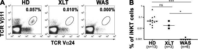
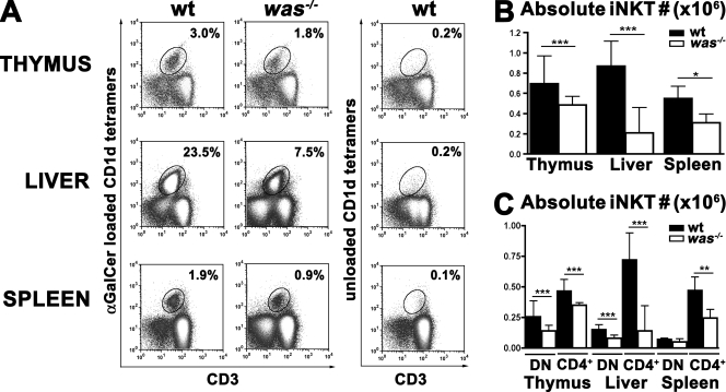
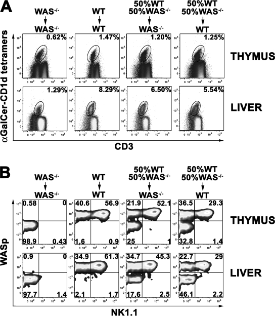
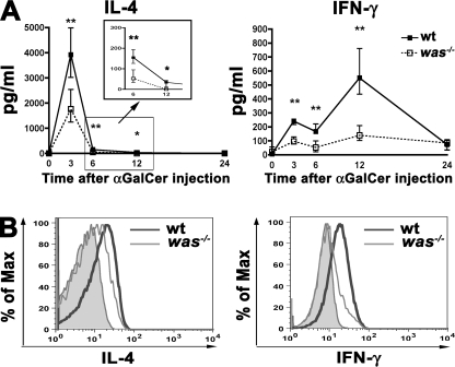
The Wiskott-Aldrich syndrome (WAS) protein (WASp) is a regulator of actin cytoskeleton in hematopoietic cells. Mutations of the WASp gene cause WAS. Although WASp is involved in various immune cell functions, its role in invariant natural killer T (iNKT) cells has never been investigated. Defects of iNKT cells could indeed contribute to several WAS features, such as recurrent infections and high tumor incidence. We found a profound reduction of circulating iNKT cells in WAS patients, directly correlating with the severity of clinical phenotype. To better characterize iNKT cell defect in the absence of WASp, we analyzed was(-/-) mice. iNKT cell numbers were significantly reduced in the thymus and periphery of was(-/-) mice as compared with wild-type controls. Moreover analysis of was(-/-) iNKT cell maturation revealed a complete arrest at the CD44(+) NK1.1(-) intermediate stage. Notably, generation of BM chimeras demonstrated a was(-/-) iNKT cell-autonomous developmental defect. was(-/-) iNKT cells were also functionally impaired, as suggested by the reduced secretion of interleukin 4 and interferon gamma upon in vivo activation. Altogether, these results demonstrate the relevance of WASp in integrating signals critical for development and functional differentiation of iNKT cells and suggest that defects in these cells may play a role in WAS pathology.


Figures

Figure 1.
Figure 1.
Lack of iNKT cells in WAS patients. (A) Representative flow cytometric analysis of peripheral blood iNKT cells from an age-matched HD control (HD), an XLT patient (XLT), and a WAS patient (WAS). Cells in the density plots are gated on CD3+ cells. Percentages of iNKT cells (TCR-Vβ11+ TCR-Vα24+) are indicated. (B) Frequency of iNKT cells from 13 controls, 3 XLT, and 6 WAS patients. Bars represent the median value of each group. ns, P > 0.05; *, P < 0.05; ***, P < 0.001.
Figure 2.
Figure 2.
Reduced iNKT cell number in was−/− mice. (A) iNKT cells were analyzed by flow cytometry in thymus, liver, and spleen of C57BL/6 WT and was−/− mice (was−/−). Thymocytes were stained with anti-CD8, anti-CD3 mAbs, and CD1d tetramers (α-GalCer loaded or unloaded), whereas hepatic leukocytes and splenocytes were stained with anti-B220 and anti-CD3 mAbs and CD1d tetramers (α-GalCer loaded or unloaded). After gating on CD8 or B220 cells, iNKT cells were identified as CD3+ CD1d tetramers+ cells. (B) Absolute numbers of iNKT cells were determined by multiplying their percentage by the absolute cell count within each sample. In A and B, data are representative of at least 10 mice per group analyzed in three independent experiments. (C) iNKT cells were stained with anti-CD4 antibodies. Comparison of the absolute number of CD4+ or CD4 (DN) iNKT cells in thymus, liver, and spleen in WT versus was−/− mice is shown. In B and C, error bars represent the median and interquartile ranges of eight mice per group. *, P < 0.05; **, P < 0.005; ***, P < 0.001.
Figure 3.
Figure 3.
Block iNKT cell maturation in the absence of WASp. (A) Thymocytes and hepatic leukocytes from WT and was−/− mice were stained with anti-CD8, anti-CD3, anti-CD44, and anti-NK1.1 mAbs and α-GalCer–loaded CD1d tetramers. Maturation of iNKT cells (CD1d tetramer+, CD3+, and CD8) was assessed by CD44 and NK1.1 expression. Data are representative of six mice per group analyzed in two independent experiments. (B) Absolute numbers of iNKT cells (CD1d tetramer+, CD3+, and CD8) in thymus and liver of WT and was−/− mice. Error bars represent median and interquartile range of six mice per group. **, P < 0.005.
Figure 4.
Figure 4.
Cell-autonomous developmental defect of was−/− iNKT cells. (A) iNKT cells were analyzed by flow cytometry in thymus and liver of WT or was−/− recipient mice (CD45.1) transplanted with was−/− lin cells, WT lin cells, or a mixture of 50% WT and 50% was−/− lin obtained from CD45.2 mice. Thymocytes and hepatic leukocytes were surface stained with α-GalCer–loaded CD1d tetramers, with anti-CD3 and anti-CD8 (thymocytes) or anti-B220 (hepatic leukocytes) mAbs. The percentage of iNKT cells (CD8 or B220 CD3+CD1d tetramer+ cells) is indicated in each plot. (B) Maturation of iNKT cells in thymus and liver of BM chimera mice. After gating on donor CD45.2+, iNKT cells were further analyzed for NK1.1 and WASp expression. The percentage of mature (NK1.1+) and immature (NK1.1) iNKT cells from WT donors (WASp+) or from was−/− donors (WASp) is indicated in each plot. Data are representative of at least three mice per group from two independent experiments.
Figure 5.
Figure 5.
Impaired cytokine production by was−/− iNKT cells. (A) In vivo IL-4 and IFN-γ production upon α-GalCer administration in WT and was−/− mice. Sera were analyzed at 3, 6, 12, and 24 h upon injection. The graphs show the amount of cytokines produced by six WT and six was−/− mice. Black and white squares represent the median values of WT and was−/− mice groups, respectively. The vertical bars represent the interquartile range of each group. *, P < 0.05; **, P < 0.005. (B) IL-4 and IFN-γ production in WT and was−/− mice at the single iNKT cell level. WT and was−/− mice were injected with α-GalCer and, after 45 min, hepatic leukocytes were isolated and stained with α-GalCer–loaded CD1d tetramers and anti-CD3, anti–IL-4, and anti–IFN-γ mAbs. Representative analysis of IL-4 and IFN-γ intracellular production by iNKT cells (CD3+ and CD1d tetramer+) from WT (thick line) and was−/− (thin line) mice is shown. Filled histograms represent IL-4 or IFN-γ production by untreated WT mice. Data are from one representative experiment of three.

References

    1. Bendelac A., Savage P.B., Teyton L. 2007. The biology of NKT cells.Annu. Rev. Immunol. 25:297–336 - PubMed
    1. Godfrey D.I., Berzins S.P. 2007. Control points in NKT-cell development.Nat. Rev. Immunol. 7:505–518 - PubMed
    1. Tupin E., Kinjo Y., Kronenberg M. 2007. The unique role of natural killer T cells in the response to microorganisms.Nat. Rev. Microbiol. 5:405–417 - PubMed
    1. Crowe N.Y., Smyth M.J., Godfrey D.I. 2002. A critical role for natural killer T cells in immunosurveillance of methylcholanthrene-induced sarcomas.J. Exp. Med. 196:119–127 - PMC - PubMed
    1. Van Kaer L. 2007. NKT cells: T lymphocytes with innate effector functions.Curr. Opin. Immunol. 19:354–364 - PubMed

Publication types