. 2009 Apr 7;119(13):1785-94.
doi: 10.1161/CIRCULATIONAHA.108.827972. Epub 2009 Mar 23.

Arterial and aortic valve calcification abolished by elastolytic cathepsin S deficiency in chronic renal disease

Affiliations

Arterial and aortic valve calcification abolished by elastolytic cathepsin S deficiency in chronic renal disease

Elena Aikawa et al. Circulation. .

Abstract

Background: Clinical studies have demonstrated that 50% of individuals with chronic renal disease (CRD) die of cardiovascular causes, including advanced calcific arterial and valvular disease; however, the mechanisms of accelerated calcification in CRD remain obscure, and no therapies can prevent disease progression. We recently demonstrated in vivo that inflammation triggers cardiovascular calcification. In vitro evidence also indicates that elastin degradation products may promote osteogenesis. Here, we used genetically modified mice and molecular imaging to test the hypothesis in vivo that cathepsin S (catS), a potent elastolytic proteinase, accelerates calcification in atherosclerotic mice with CRD induced by 5/6 nephrectomy.

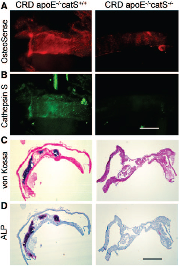
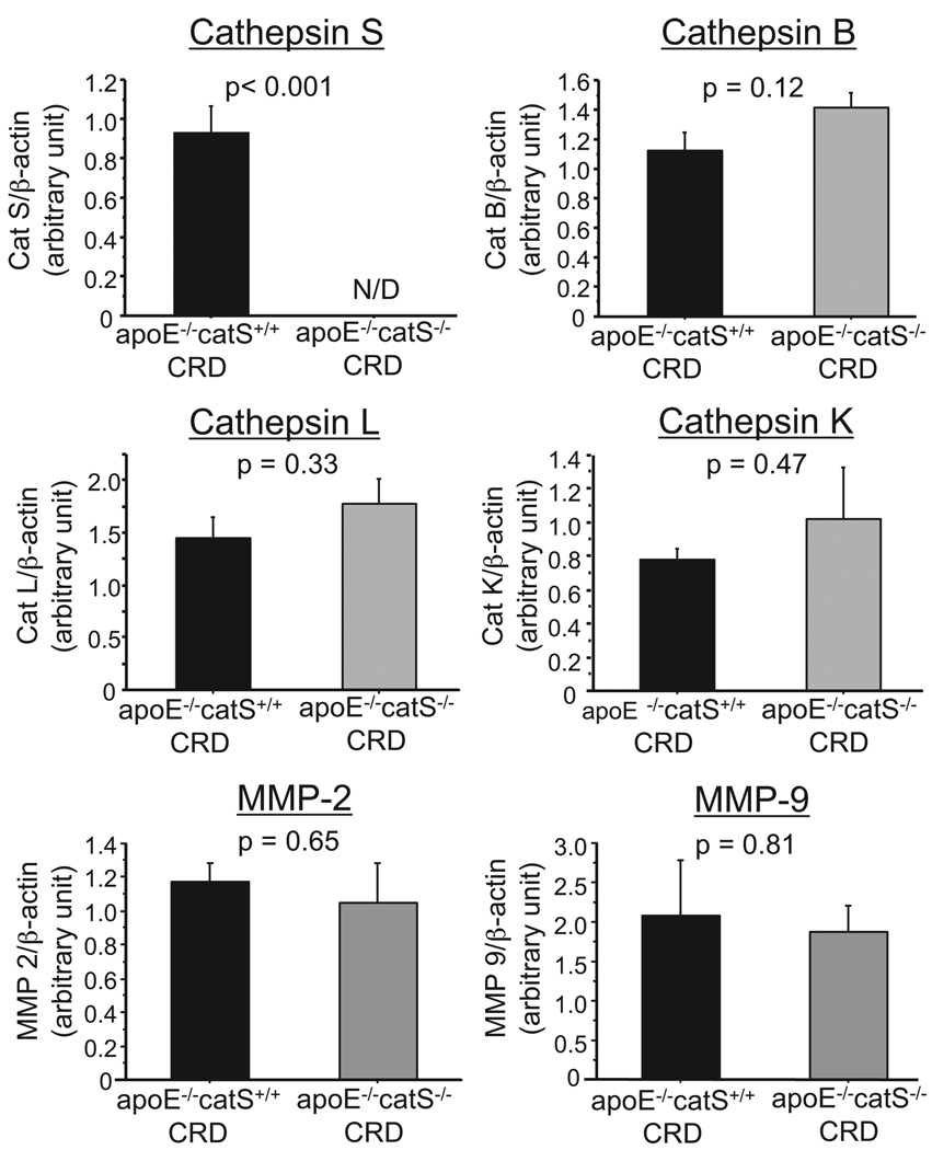
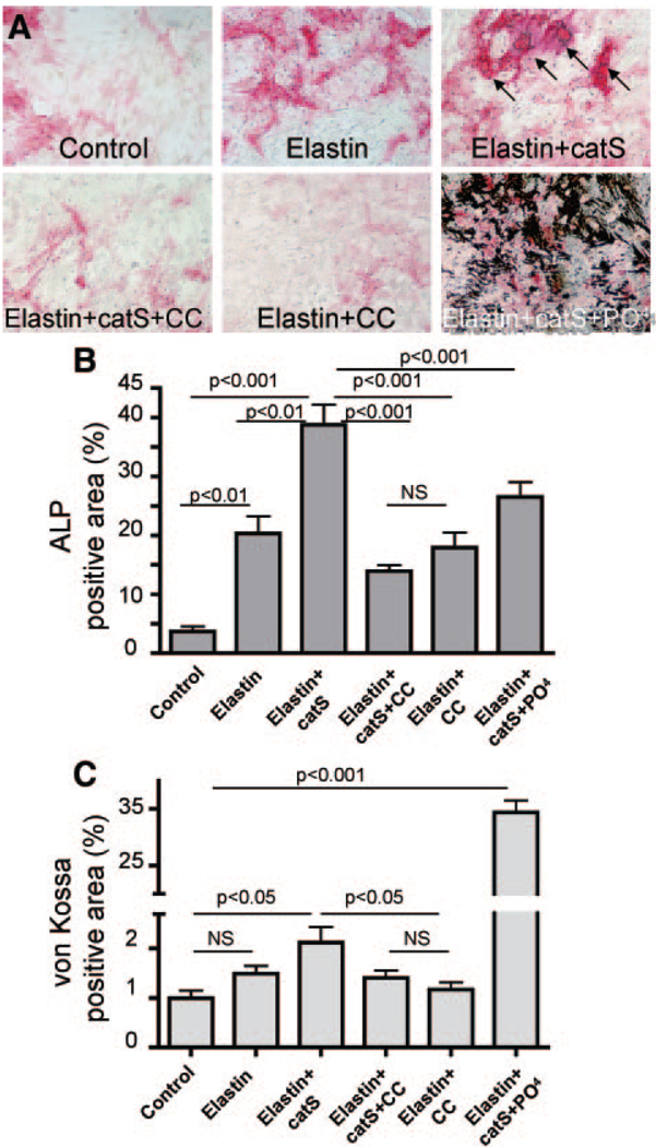
Methods and results: Apolipoprotein-deficient (apoE(-/-))/catS(+/+) (n=24) and apoE(-/-)/catS(-/-) (n=24) mice were assigned to CRD and control groups. CRD mice had significantly higher serum phosphate, creatinine, and cystatin C levels than those without CRD. To visualize catS activity and osteogenesis in vivo, we coadministered catS-activatable and calcification-targeted molecular imaging agents 10 weeks after nephrectomy. Imaging coregistered increased catS and osteogenic activities in the CRD apoE(-/-)/catS(+/+) cohort, whereas CRD apoE(-/-)/catS(-/-) mice exhibited less calcification. Quantitative histology demonstrated greater catS-associated elastin fragmentation and calcification in CRD apoE(-/-)/catS(+/+) than CRD apoE(-/-)/catS(-/-) aortas and aortic valves. Notably, catS deletion did not cause compensatory increases in RNA levels of other elastolytic cathepsins or matrix metalloproteinases. Elastin peptide and recombinant catS significantly increased calcification in smooth muscle cells in vitro, a process further amplified in phosphate-enriched culture medium.

Conclusions: The present study provides direct in vivo evidence that catS-induced elastolysis accelerates arterial and aortic valve calcification in CRD, providing new insight into the pathophysiology of cardiovascular calcification.


Figures

Figure 1
Figure 1
Calcification increased in aortas of CRD apoE−/−/catS+/+ mice and decreased with catS deletion. A, Macroscopic fluorescence reflectance imaging of calcification in CRD apoE−/−/catS+/+ mice yielded strong osteogenic signals in the entire aorta compared with aortas of CRD apoE−/−/catS−/− mice and mice with normal renal function. Bar = 1 cm. Histological analysis (B; von Kossa; bar = 200 µm) and quantitative assessment (C through E) showed that only CRD apoE−/−/catS+/+ mice had increased calcification, whereas no significant difference in plaque size and macrophage (Mac-3) accumulation was observed between CRD apoE−/−/catS+/+ and CRD apoE−/−/catS−/− mice.
Figure 2
Figure 2
Increased osteogenic action and catS activity in carotid atherosclerotic plaques of CRD apoE−/−/catS+/+ mice. A and B, Intravital microscopy revealed increased osteogenic and catS signal intensity in CRD apoE−/−/catS+/+ mice vs lower osteogenic signal and negligible elastolytic catS activity in CRD apoE−/−/catS−/−. Bar = 500 µm. C and D, Correlative histological analysis showed advanced calcification in CRD apoE−/−/catS+/+ mouse artery (von Kossa and alkaline phosphatase activity [ALP]), whereas CRD apoE−/−/catS−/− mice exhibited no calcification. Bar = 200 µm.
Figure 3
Figure 3
Increased catS-associated elastin fragmentation in CRD apoE−/−/catS+/+ mouse aorta. A, Fluorescence immunohistochemistry detected abundant catS expression (red) in CRD apoE−/−/catS+/+ plaque associated with increased fragmentation of elastin (green). apoE−/−/catS−/− and CRD apoE−/−/catS−/− mice had negligible catS expression and a reduced number of elastin fragments, confirmed by quantitative histological assessment (B). L indicates lumen. Dotted line depicts the plaque margin. Bar = 50 µm.
Figure 4
Figure 4
Intimal and medial calcification was associated with catS expression and elastin degradation in CRD apoE−/−/catS+/+ mouse aortas. A, Histological analysis revealed distinct phenotypic changes in the atheroma of CRD apoE−/−/catS+/+ and CRD apoE−/−/catS−/− mice: Advanced calcification in the intima and scarce in the media (von Kossa; arrows) of CRD apoE−/−/catS+/+ mouse aorta was associated with elastin loss (van Gieson; arrowheads), whereas CRD apoE−/−/catS−/− mice had preserved elastin and undetectable calcification. B, Adjacent sections stained with hematoxylin and eosin (HE) showed an association of catS expression with calcifying cells in the media and intima (ALP [alkaline phosphatase] and von Kossa; arrows) and insufficient elastin (van Gieson; arrowheads) in CRD apoE−/−/catS+/+ plaque. Notably, cystatin C tissue expression was considerably low. C, Fluorescence imaging colocalized immunoreactive catS (green) with osteogenic near-infrared signal (OsteoSense680; red). Calcifying cells (yellow) surrounded by fragmented elastin fibers (arrows) were evident on the merged image. Bars = 50 µm.
Figure 5
Figure 5
Inflamed aortic valves of CRD apoE−/−/catS+/+ mice had characteristics of early calcific disease. A, Gross morphology and ex vivo fluorescence microscopy (image stacks) of calcified aortic valves (top) visualized osteogenic activity (OsteoSense, red) in the areas of leaflet attachment to the aortic wall in inflamed valves (CLIO-gly, green). Individual mapping (bottom) showed maximum signal intensities at the areas of high mechanical stress (arrows). The data represent 3 mice that showed similar results. B, Aortic leaflets from CRD apoE−/−/catS+/+ mice contained abundant macrophages (Mac 3) that expressed catS in association with undetectable elastin (van Gieson negative) and showed evidence of calcification (von Kossa), the characteristics of calcific aortic valve disease (left). In contrast, inflamed leaflets of CRD apoE−/−/catS−/− mice exhibited negligible catS expression, preserved elastin, and no evidence of calcification (right). Bar = 100 µm. C, Quantitative assessment showed no significant difference in macrophage accumulation between CRD apoE−/−/catS+/+ and CRD apoE−/−/catS−/− mice. Ao indicates aorta.
Figure 6
Figure 6
CatS deletion did not alter expression levels of major elastolytic enzymes. Quantitative real-time reverse-transcription polymerase chain reaction demonstrated β-actin–adjusted levels of RNAs encoding cathepsins S, B, L, and K and MMP-2 and MMP-9 in the aortas of CRD apoE−/−/catS+/+ or CRD apoE−/−/catS−/− mice. CRD apoE−/−/catS−/− mouse aortas did not express detectable levels of catS. CatS deletion did not cause statistically significant changes in expression of elastolytic cathepsins and MMPs.
Figure 7
Figure 7
Elastin degradation and high phosphate levels promote vascular SMC calcification. A, Human primary vascular SMCs were treated with elastin peptides (elastin), alone or in the presence of catS, CatS plus cathepsin inhibitor cystatin C (CC), or catS plus sodium monophosphate (PO4), as shown. After treatment, cells were stained for alkaline phosphatase (ALP) activity and then fixed and restained for the presence of calcium phosphate crystals with von Kossa. B and C, ALP activity (B; red reaction product) and (C) hydroxyapatite crystals stained with von Kossa (black) were quantified as percentage of total area.
Figure 8
Figure 8
Schematic of the proposed multistep mechanism of accelerated atherosclerotic and valvular calcification in CRD. In atherosclerotic artery and inflamed aortic valve, macrophage-derived catS degrades elastin. Biologically active elastin-degradation products initiate calcification of mesenchymal cells (eg, vascular SMCs or valvular myofibroblast-like cells). Furthermore, CRD-associated hyperphosphatemia accelerates calcification of mesenchymal cells through phosphate-induced release of matrix vesicles and apoptosis, which results in more advanced calcification. PO4 indicates sodium monophosphate.

References

    1. Otto CM. Calcific aortic stenosis: time to look more closely at the valve. N Engl J Med. 2008;359:1395–1398. - PubMed
    1. Johnson RC, Leopold JA, Loscalzo J. Vascular calcification: pathobiological mechanisms and clinical implications. Circ Res. 2006;99:1044–1059. - PubMed
    1. Demer LL, Tintut Y. Vascular calcification: pathobiology of a multifaceted disease. Circulation. 2008;117:2938–2948. - PMC - PubMed
    1. Towler DA. Vascular calcification: a perspective on an imminent disease epidemic. IBMS BoneKEy. 2008;5:41–58.
    1. Fox CS, Larson MG, Vasan RS, Guo CY, Parise H, Levy D, Leip EP, O’Donnell CJ, D’Agostino RB, Sr, Benjamin EJ. Cross-sectional association of kidney function with valvular and annular calcification: the Framingham Heart Study. J Am Soc Nephrol. 2006;17:521–527. - PubMed

Publication types

MeSH terms