

doi: 10.1073/pnas.0809483106.
Epub 2009 Mar 23.
Adult deafness induces somatosensory conversion of ferret auditory cortex
Affiliations
- PMID: 19307553
- PMCID: PMC2667075
- DOI: 10.1073/pnas.0809483106
Item in Clipboard
Adult deafness induces somatosensory conversion of ferret auditory cortex
Proc Natl Acad Sci U S A.
.
Abstract
In response to early or developmental lesions, responsiveness of sensory cortex can be converted from the deprived modality to that of the remaining sensory systems. However, little is known about capacity of the adult cortex for cross-modal reorganization. The present study examined the auditory cortices of animals deafened as adults, and observed an extensive somatosensory conversion within as little as 16 days after deafening. These results demonstrate that cortical cross-modal reorganization can occur after the period of sensory system maturation.
Conflict of interest statement
The authors declare no conflict of interest.
Figures
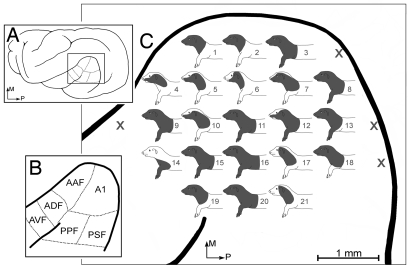
Cross-modal reorganization of auditory cortex in a ferret deafened as an adult. (A) The location of the auditory cortex (box) on lateral view of the ferret brain; (B) auditory cortical areas (after 32). (C) An enlarged view of the auditory cortices in an adult-deafened ferret (175 DPN at deafening, 74 d deaf) shows each recording site indicated by the numbered somatosensory receptive field that was mapped at that site (X, unresponsive). All auditory areas tested [corresponding to A1, anterior auditory field (AAF), and portions of anterior dorsal field (ADF) and posterior pseudo-sylvian field (PPF)] were responsive to somatosensory stimulation, primarily on the head. All recordings were made at 1,000 μm depth. (AVF, anterior ventral field; PSF, posterior suprasylvian field.)
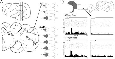
Somatosensory responses from auditory cortex in adult-deafened ferrets. (A). The lateral view of the ferret brain shows the location of the sections (Bottom Left) containing the recording penetrations in areas A1 and anterior auditory field (AAF) from an adult ferret 64 days post-deafening. Recording penetrations sampled neurons at 250 μm intervals (for space reasons, the examples shown are at 500-μm intervals) with corresponding receptive fields indicated on ferret body plots. For each penetration, receptive fields were on the head and were uniformly bilateral, and somatosensory responses spanned the full cortical thickness. (B) Recordings made from an adult ferret at 16 days post-deafening. Two representative neurons (950 and 1,150 μm deep) from area A1 (marked on lateral view of brain) exhibited somatosensory receptive fields on the head, neck, and forelimb. The peristimulus-time histograms show that repeated, electronically triggered tactile stimulation within the receptive field (cheek) evoked vigorous responses in both neurons, but stimulation outside the receptive field (torso) failed to elicit responses. (Tactile stimulus, ramp labeled S; raster 1 dot represents 1 spike; each row represents 1 trial; histogram, 10-ms time bins.)
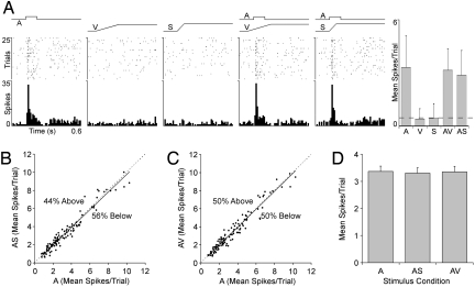
Responses of auditory neurons from hearing adult ferret to auditory, visual, somatosensory, and multi-sensory stimulation. (A) Responses of a typical neuron in A1 of a hearing adult to auditory (square-wave labeled A, contralateral white noise, 75 dB SPL, 100 ms), visual (ramp labeled V, light bar moved across the visual field), somatosensory (ramp labeled S, tactile probe indented skin on contralateral cheek), and combined stimulation (AV and AS) are shown in the rasters (1 dot represents 1 spike; each row represents 1 trial) and histograms (10-ms time bins). As summarized in the bar graph (mean spikes/trial ± SD; spontaneous activity indicated by dashed line), the neuron responded vigorously to the auditory stimulus, with no response to the visual or somatosensory stimulus. Because the response to the auditory stimulus alone was not statistically different from that of the combined stimuli conditions (AV or AS), this auditory neuron did not show subthreshold cross-modal effects. For the population of auditory neurons examined (n = 111), B and C plot the responses to the auditory stimulus alone (A; x axis) to those evoked by the combined stimuli (AS or AV; y axis). In both graphs, most responses fell on or near the line of unity (dashed), with similar numbers either slightly above or below. (D) Bar graph shows the population response average (mean spikes/trial ± SEM) to the auditory stimulus alone (A), auditory-somatosensory combined stimuli (AS), and auditory-visual combined stimuli (AV), which were not significantly different (P > 0.05).
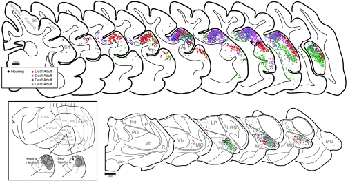
Anatomical tracer injected into A1 revealed the same pattern of connections in both adult-deafened (≈76 d duration) and hearing ferrets. (Inset) Lateral view of ferret cortex with expanded views of the auditory cortices depicting the tracer deposits (gray) for hearing and deafened animals. (Top) Serially arranged coronal sections containing somatosensory and auditory cortices. Each dot represents 1 labeled neuron (black dots represent 1 hearing adult ferret; colored dots represent 4 deafened adult ferrets). The distribution of black and colored dots is essentially co-extensive; areas of somatosensory cortex (S1; SII) are largely devoid of label. No labeled neurons were identified in sections anterior or posterior to those depicted. (Bottom) Serially arranged sections through thalamus with the auditory (MG, medial geniculate), somatosensory (Vb, ventrobasal), visual (LGN, lateral geniculate; Pul, pulvinar), and non-specific (PO, posterior; LP, lateral posterior) nuclei depicted. Each dot represents 1 labeled neuron (black dots, hearing; colored dots, deafened). The distribution of black and colored dots is essentially co-extensive within the MG; somatosensory thalamus (Vb) is devoid of label in both conditions.
References
-
- Morishita H, Hensch TK. Critical period revisited: impact on vision. Curr Opin Neurobiol. 2008;18:101–107. - PubMed
-
- Hyvarinen J, Carlson S, Hyvarinen L. Early visual deprivation alters modality of neuronal responses in area 19 of monkey cortex. Neurosci Lett. 1981;26:239–243. - PubMed
-
- Izraeli R, et al. Cross-modal neuroplasticity in neonatally enucleated hamsters: structure, electrophysiology and behaviour. Eur J Neurosci. 2002;15:693–712. - PubMed
Publication types
MeSH terms
Grants and funding
LinkOut - more resources
Full Text Sources
Medical
