. 2009 Apr 7;106(14):5546-50.
doi: 10.1073/pnas.0901715106. Epub 2009 Mar 23.

Nanotechnology approach for drug addiction therapy: gene silencing using delivery of gold nanorod-siRNA nanoplex in dopaminergic neurons

Affiliations

Nanotechnology approach for drug addiction therapy: gene silencing using delivery of gold nanorod-siRNA nanoplex in dopaminergic neurons

Adela C Bonoiu et al. Proc Natl Acad Sci U S A. .

Abstract

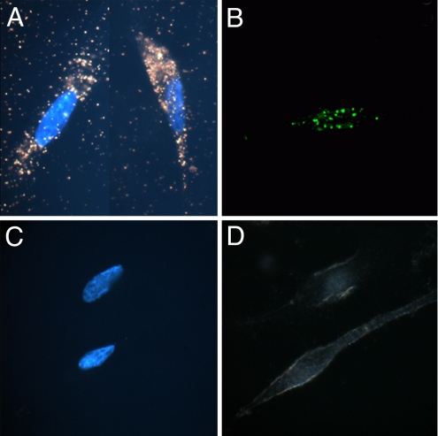
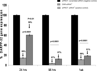
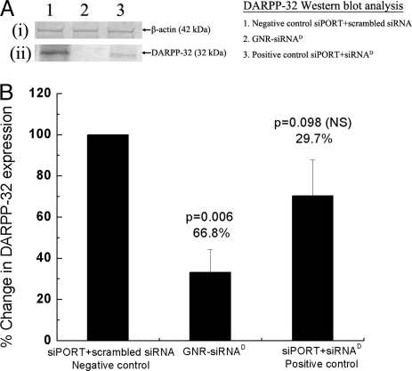
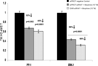
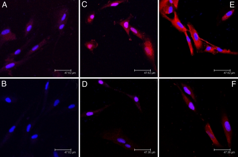
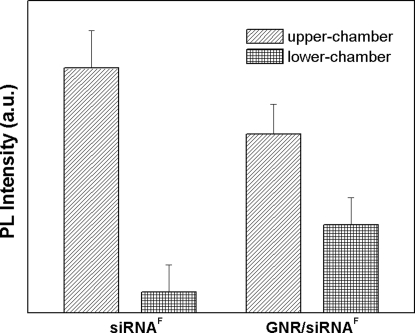
Drug abuse is a worldwide health concern in which addiction involves activation of the dopaminergic signaling pathway in the brain. Here, we introduce a nanotechnology approach that utilizes gold nanorod-DARPP-32 siRNA complexes (nanoplexes) that target this dopaminergic signaling pathway in the brain. The shift in the localized longitudinal plasmon resonance peak of gold nanorods (GNRs) was used to show their interaction with siRNA. Plasmonic enhanced dark field imaging was used to visualize the uptake of these nanoplexes in dopaminergic neurons in vitro. Gene silencing of the nanoplexes in these cells was evidenced by the reduction in the expression of key proteins (DARPP-32, ERK, and PP-1) belonging to this pathway, with no observed cytotoxicity. Moreover, these nanoplexes were shown to transmigrate across an in vitro model of the blood-brain barrier (BBB). Therefore, these nanoplexes appear to be suited for brain-specific delivery of appropriate siRNA for therapy of drug addiction and other brain diseases.


Conflict of interest statement

The authors declare no conflict of interest.

Figures

Fig. 1.
Fig. 1.
Study of GNR-siRNA nanoplex uptake in DAN cells. Dark-field images with Hoechst nuclear staining (A and C), and confocal images (B and D) of DAN cells, following treatment with GNR-siRNA nanoplex (A and B) and free siRNA (C and D). The dark-field images of GNRs corresponding to the longitudinal surface plasmonic enhancement in the red region can be clearly distinguished from the background (A). Confocal images show robust uptake of the siRNAF complexed with GNRs (B), as opposed to free siRNAF (C).
Fig. 2.
Fig. 2.
DARPP-32 gene silencing efficiency of GNR-siRNAD nanoplex in DAN cells. Quantitative real-time PCR (Q-PCR) data showing a >80% suppression of DARPP-32 gene expression in DAN cells that were transfected with the GNR-siRNAD nanoplex. Relative expression of mRNA species was calculated using the comparative CT method. Data are the mean ± SD of 3 separate experiments done in duplicate. Statistical significance was determined using ANOVA based on comparison between the siPORT-siRNAD (positive control), GNR-siRNAD nanoplex, and the siPORT with scrambled siRNA (negative control). A significant decrease in DARPP-32 gene expression was observed up to 1 week posttransfection, with maximum suppression observed at 96 h posttransfection.
Fig. 3.
Fig. 3.
Effect of the GNR-siRNAD nanoplex on DARPP-32 protein expression in DAN cells. Western blot analysis of DARPP-32 protein from lysates of DAN cells following their treatment in vitro with GNR-siRNAD nanoplexes (lane 2) and both siPORT positive control (lane 3) and negative control (lane 1), at 120 h posttransfection. Panel A shows data from a representative Western blot experiment showing (i) no change in b-actin (42-kDa band) protein expression and (ii) a significant decrease in DARPP-32 (32-kDa band) protein expression, in cells treated with the GNR-siRNAD nanoplex (lane 2), as opposed to cells treated with negative control (siPORT-scrambled siRNA, lane 1). Panel B is a graphical representation of the densitometric analysis of the DARPP-32 protein band showing percentage decrease in the DARPP-32 protein expression in cells treated with GNR-siRNAD and siPORT-siRNAD, when compared with that in cells treated with siPORT-scrambled siRNA (negative control, 100% DARPP-32 expression). Data shown are mean ± SD of results from 3 separate experiments, and statistical significance was determined using ANOVA. Results show a significant decrease in DARPP-32 protein expression in the GNR-siRNAD-treated cells (66.8% decrease), significantly higher than that obtained from siPORT-siRNAD-treated cells (29.7% decrease).
Fig. 4.
Fig. 4.
Modulation of gene expression of effector molecules downstream of DARPP-32 (PP-1 and ERK-1) in siRNAD transfected DAN cells. Q-PCR data show a significant decrease in both PP-1 and ERK-1 gene expression in DAN cells following treatment with GNR-siRNAD nanoplexes. Data are the mean ± SD of 3 separate experiments performed in duplicate. Statistical significance was determined using ANOVA based on comparison between the GNR-siRNAD and siPORT- siRNAD (positive control) samples with the siPORT-scrambled siRNA (negative control).
Fig. 5.
Fig. 5.
Immunofluorescent studies on neuronal cells. Immunofluorescent staining for DARPP-32 (A–D) and ERK-1 (E and F) in transfected and nontransfected DAN cells. (A) DARPP-32 expression in nontransfected DAN cells. (B) DARPP-32 expression in GNR-siRNAD nanoplex transfected DAN cells. (C) Morphine-treated (10−7 M) but nontransfected DAN cells. (D) Morphine-treated (10−7 M) DAN cells, transfected with GNR-siRNAD. (E) Morphine-treated (10−7 M) untransfected DAN cells showing ERK-1 expression. (F) Morphine-treated (10−7 M) GNR-siRNAD tranfected DAN cells showing ERK-1 expression. Our results show (A) morphine treatment enhances the DARPP-32 levels, and (B) GNR-mediated transfection of siRNA against DARPP-32 reduces not only the levels of DARPP-32 expression but also the expression of its downstream effector molecule ERK-1 in DAN cells.
Fig. 6.
Fig. 6.
Transmigration of GNR-siRNAF nanoplex across the brain blood–barrier model. Fluorescence signal distribution collected from BBB model in upper chamber (blood end) and lower chamber (brain end) of an in vitro BBB model, following treatment with free siRNAF and GNR-siRNAF nanoplex. Results show enhanced transmigration efficiency of the GNR-siRNAF nanoplex when compared to that of free siRNAF.

References

    1. Prasad PN. Introduction to Biophotonics. New York: Wiley; 2004.
    1. Kang H, DeLong R, Fisher MH, Juliano R. Tat-conjugated PAMAM dendrimers as delivey agents for antisense and siRNA oligonucleotides. J Pharm Res. 2005;22(12):2099–2106. - PubMed
    1. Farokhzad OC, Langer R. Nanomedicine: Developing smarter therapeutic and diagnostic modalities. Adv Drug Delivery Rev. 2006;58(14):1456–1459. - PubMed
    1. Pardridge WM. Re-engineering biopharmaceuticals for delivery to brain with molecular Trojan horses. Bioconjug Chem. 2008;19(7):1327–1338. - PubMed
    1. Ferrari M. Cancer nanotechnology: Opportunities and challenges. Nat Rev Cancer. 2005;5(3):161–171. - PubMed

Publication types

MeSH terms