. 2009 Apr 7;106(14):5784-8.
doi: 10.1073/pnas.0901425106. Epub 2009 Mar 23.

Rational development of high-affinity T-cell receptor-like antibodies

Affiliations

Rational development of high-affinity T-cell receptor-like antibodies

Guillaume Stewart-Jones et al. Proc Natl Acad Sci U S A. .

Erratum in

  • Proc Natl Acad Sci U S A. 2009 Jun 30;106(26):10872. Nuber, Natko [added]

Abstract

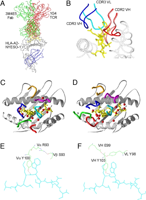
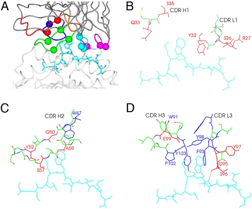
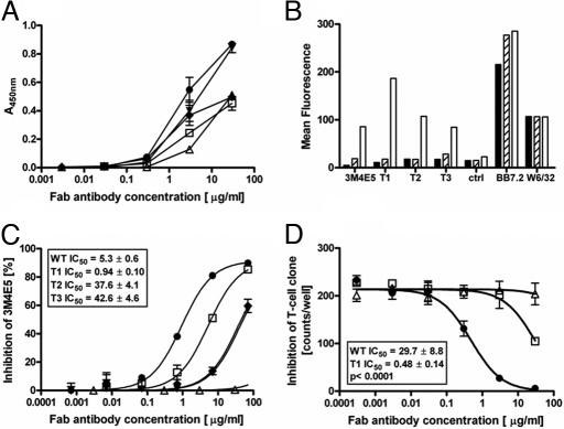
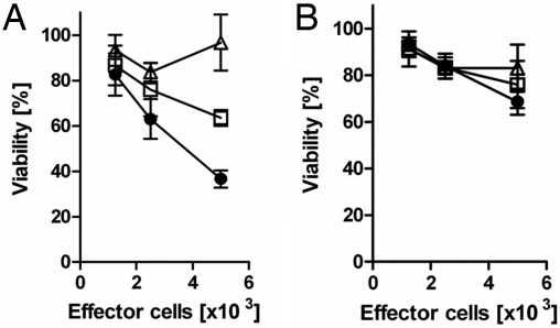
T-cell interaction with a target cell is a key event in the adaptive immune response and primarily driven by T-cell receptor (TCR) recognition of peptide-MHC (pMHC) complexes. TCR avidity for a given pMHC is determined by number of MHC molecules, availability of coreceptors, and TCR affinity for MHC or peptide, respectively, with peptide recognition being the most important factor to confer target specificity. Here we present high-resolution crystal structures of 2 Fab antibodies in complex with the immunodominant NY-ESO-1(157-165) peptide analogue (SLLMWITQV) presented by HLA-A*0201 and compare them with a TCR recognizing the same pMHC. Binding to the central methionine-tryptophan peptide motif and orientation of binding were almost identical for Fabs and TCR. As the MW "peg" dominates the contacts between Fab and peptide, we estimated the contributions of individual amino acids between the Fab and peptide to provide the rational basis for a peptide-focused second-generation, high-affinity antibody library. The final Fab candidate achieved better peptide binding by 2 light-chain mutations, giving a 20-fold affinity improvement to 2-4 nM, exceeding the affinity of the TCR by 1,000-fold. The high-affinity Fab when grafted as recombinant TCR on T cells conferred specific killing of HLA-A*0201/NY-ESO-1(157-165) target cells. In summary, we prove that affinity maturation of antibodies mimicking a TCR is possible and provide a strategy for engineering high-affinity antibodies that can be used in targeting specific pMHC complexes for diagnostic and therapeutic purposes.


Conflict of interest statement

The authors declare no conflict of interest.

Figures

Fig. 1.
Fig. 1.
Structural comparison of TCR and Fab. (A) Comparison of positions of 1G4 TCR (red) and 3M4E5 Fab in complex with HLA-A*0201/NY-ESO-1157–167. (B) Positions of the CDR3 loops (blue and magenta) bound to the peptide (cyan) and MHC (gray), and the 2 positions of the CDR2 VH loop from the 3M4F4 complex in the up (red) and down conformations. (C) Schematic of CDR loops of 1G4 TCR in complex with HLA-A*0201/NY-ESO-1157–167 with loops colored as follows: Vα CDR1, green; Vα CDR2, red; Vα CDR3, blue; Vβ CDR1, magenta; Vβ CDR2, orange; and Vβ CDR3, cyan. Water molecules within 4 Å of both TCR and peptide are indicated as red spheres. (D) Diagram of CDR loops of 3M4E5 Fab in complex with HLA- A*0201/NYESO-1157–167 with loops colored as follows: VH CDR1, green; VH CDR2, red; VH CDR3, blue; VL CDR1, magenta; VL CDR2, orange; and VL CDR3, cyan. Water molecules within 4 Å of both TCR and peptide are indicated as red spheres. (E) Structure of the 1G4 TCR residues forming the “roof” residues in the cavity that binds the peptide (cyan) MW side chains. (F) The analogous 3M4E5 Fab side chains that form the peptide MW binding cavity. Hydrogen bonds are shown in black dashed lines.
Fig. 2.
Fig. 2.
Structural basis for a second-generation library. (A) Side view of 3M4E5 Fab in complex with HLA-A*0201/NY-ESO-1157–167, with residues randomized in the second-generation phage display library highlighted as spheres. Colors are as in Fig. 1D. (B–D) Illustrations of 3M4E5 Fab CDR residues that were randomized (red) in the vicinity of the peptide, which displayed nonoptimal contacts with the peptide. Residues making key contacts with the peptide MW side chains and lining the cavity in the CDR loops that interacts with the MW peg are colored in blue. The NYESO-1157–167 peptide is colored cyan, and hydrogen bonds are represented as dashed black lines.
Fig. 3.
Fig. 3.
Binding characteristics of 3M4E5-evolved Fab antibodies T1–3. (A) Comparison of binding characteristics as detected by ELISA on HLA-A*0201/NY-ESO-1157–165 complexes. Fab type 1 T1 (filled circles), type 2 T2 (diamonds), type 3 T3 (filled triangles), control Fab (open triangles) with weak binding characteristics, and Fab WT 3M4E5 (open squares) were titrated over the indicated concentration range. (B) Confirmation of Fab binding specificity was achieved by incubation with minigene transfected T2 cells. Reactivity of 3M4E5 and Fabs T1–T3 molecules with target HLA-A*0201/NY-ESO-1157–165 complex (white column) and control complexes (black HLA-A*0201/NY-ESO-1157–167 and gray HLA-A*0201/NY-ESO-1155–163) is depicted. HLA-A*0201 expression was confirmed for all cell lines by BB7.2 and w6/32 antibody staining, respectively. (C) Inhibition of 3M4E5-b binding by competition FACS analysis. Coincubation of PE-labeled biotinylated 3M4E5-b tetramers and different concentrations (50, 5, 0.5, 0.05, 0.005 0.0005, 0 μg/mL) of nonlabeled 3M4E5 (open squares), T1 (filled circles), T2 (filled triangles), T3 (filled triangles), or control Fab (open triangles) with peptide-pulsed T2 cells. (D) Inhibition of NY-ESO-1157–165-specific CTL responses by indicated Fab concentrations of T1 (filled circles), Fab 3M4E5 (open squares), or irrelevant Fab (open triangles). IFN-γ response of NY-ESO-1157–165-specific CTL to T2 cells pulsed with SLLMWITQC peptide (10−6 M) was detected by ELISPOT. All assays were done in triplicate.
Fig. 4.
Fig. 4.
Specific lysis of HLA-A*0201-positive T2 cells by recombinant immunoreceptors. CD3+ T cells were grafted by retroviral gene transfer with different recombinant immunoreceptors [T1 (filled circles), 3M4E5 (open squares), anti-CEA scTCR (open triangles)] and cocultivated (1.25–10 × 103/well) with HLA-A*0201-positive T2 cells (5 × 10⁴/well) expressing either the NY-ESO-1157–165 (A) or IMP58–66 Flu peptide (B). Viability of target cells was recorded by a XTT-based viability assay as described in Materials and Methods. All assays were performed at least in triplicate with T1 always achieving a significantly (P < 0.05) higher cytotoxic activity.

Similar articles

Cited by

References

    1. Mak TW. The T cell antigen receptor: “The Hunting of the Snark.”. Eur J Immunol. 2007;37(Suppl 1):S83–S93. - PubMed
    1. Chames P, Hufton SE, Coulie PG, Uchanska-Ziegler B, Hoogenboom HR. Direct selection of a human antibody fragment directed against the tumor T-cell epitope HLA-A1-MAGE-A1 from a nonimmunized phage-Fab library. Proc Natl Acad Sci USA. 2000;97(14):7969–7974. - PMC - PubMed
    1. Konig R. Interactions between MHC molecules and co-receptors of the TCR. Curr Opin Immunol. 2002;14(1):75–83. - PubMed
    1. Zhao Y, et al. High-affinity TCRs generated by phage display provide CD4+ T cells with the ability to recognize and kill tumor cell lines. J Immunol. 2007;179(9):5845–5854. - PMC - PubMed
    1. Holler PD, Chlewicki LK, Kranz DM. TCRs with high affinity for foreign pMHC show self-reactivity. Nat Immunol. 2003;4(1):55–62. - PubMed

Publication types

MeSH terms