. 2009 Mar 23;184(6):817-31.
doi: 10.1083/jcb.200809028.

Regulation of epithelial-mesenchymal IL-1 signaling by PPARbeta/delta is essential for skin homeostasis and wound healing

Affiliations

Regulation of epithelial-mesenchymal IL-1 signaling by PPARbeta/delta is essential for skin homeostasis and wound healing

Han Chung Chong et al. J Cell Biol. .

Abstract

Skin morphogenesis, maintenance, and healing after wounding require complex epithelial-mesenchymal interactions. In this study, we show that for skin homeostasis, interleukin-1 (IL-1) produced by keratinocytes activates peroxisome proliferator-activated receptor beta/delta (PPARbeta/delta) expression in underlying fibroblasts, which in turn inhibits the mitotic activity of keratinocytes via inhibition of the IL-1 signaling pathway. In fact, PPARbeta/delta stimulates production of the secreted IL-1 receptor antagonist, which leads to an autocrine decrease in IL-1 signaling pathways and consequently decreases production of secreted mitogenic factors by the fibroblasts. This fibroblast PPARbeta/delta regulation of the IL-1 signaling is required for proper wound healing and can regulate tumor as well as normal human keratinocyte cell proliferation. Together, these findings provide evidence for a novel homeostatic control of keratinocyte proliferation and differentiation mediated via PPARbeta/delta regulation in dermal fibroblasts of IL-1 signaling. Given the ubiquitous expression of PPARbeta/delta, other epithelial-mesenchymal interactions may also be regulated in a similar manner.


Figures
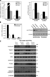
Figure 1.
Figure 1.
PPARβ/δ-deficient fibroblasts increase epidermal proliferation. (A) Expression profile of PPARs in OTC keratinocytes and fibroblasts. Total RNA and protein were extracted from keratinocytes and fibroblasts in OTC. Expression levels of PPAR mRNA (left) and protein (right) were monitored by qPCR and PPAR transcription factor assay kit, respectively. PPARβ/δ mRNA was normalized with control ribosomal protein P0 mRNA. (B) Human keratinocytes or fibroblasts were transduced with a lentiviral vector harboring a control or two different PPARβ/δ (PPARβ/δ9 and PPARβ/δ11) siRNAs. (C) Immunoblot analysis of epidermis from 2-wk-old OTCs constructed using KCTRL or KPPARβ/δ and FCTRL or FPPARβ/δ. Involucrin and transglutaminase I (Tgase I) are terminal differentiation markers, and keratin 10 (CK10) is an early differentiation marker. Keratin 5 (CK5) identifies the basal keratinocytes. Cell proliferation was measured using PCNA and cyclin D1. Apoptosis was detected using caspase 3. β-Tubulin showed equal loading and transfer. Representative immunoblots of epidermis from two OTCs are shown. Data are mean ± SD, n = 3.
Figure 2.
Figure 2.
Reduced fibroblast PPARβ/δ expression increases IL-1β activation of TAK1 and up-regulation of AP-1–controlled mitogenic target genes. (A) Expression of mitogenic factor mRNAs in 2-wk old OTC FPPARβ/δ and FCTRL treated with PPARβ/δ agonist (500 nM GW501516 for 24 h) or vehicle. The expression levels of the indicated mitogenic factors were analyzed by qPCR and normalized to control ribosomal protein P0. Results are represented in fold induction as compared with OTC FCTRL. (B) Immunoblot analysis of phosphorylated c-Jun from FPPARβ/δ and FCTRL extracted from KCTRL/FPPARβ/δ and KCTRL/FCTRL OTCs (n = 2), respectively. Total c-Jun and TAK1 protein expression level, which remains unchanged, showed equal loading and transfer. (C) Immunoblot analysis of IL-1β and TNF-α activation of TAK1 in FCTRL or FPPARβ/δ. Cells were treated with either vehicle (DMSO) or 800 nM GW501516 for 24 h prior to exposure to 10 ng/ml IL-1β (top) or TNF-α (bottom). At the indicated time points, total cell lysates were extracted. Equal amounts of total protein (50 µg) were resolved, electrotransferred, and probed for phosphorylated TAK1 (Thr184/187), total TAK1, and β-tubulin. Values below each band represent the mean fold differences (n = 3) in expression level with respect to vehicle-treated FCTRL at 5 min, which was assigned the value of one. Data are mean ± SEM, n = 3.
Figure 3.
Figure 3.
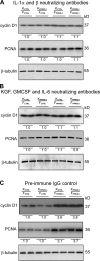
Neutralizing antibodies against IL-1α/β or KGF, GMCSF, and IL-6 abolished the mitogenic effect of FPPARβ/δ. (A–C) Immunoblot analysis of epidermis from indicated OTCs treated with 400 ng/ml IL-1α/β (A), KGF, GMCSF, and IL-6 (B) neutralizing antibodies (each at 400 ng/ml) or 400 ng/ml preimmune IgG (C). Antibodies were added to OTC medium at each change of medium. Cell proliferation was measured using cyclin D1 and PCNA. Values below each band represent the mean fold differences in expression level with respect to KCTRL from KCTRL/FCTRL OTC, which was assigned the value of 1. β-Tubulin served as a loading control. Representative immunoblots of epidermis from two indicated OTCs are shown.
Figure 4.
Figure 4.
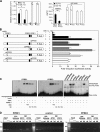
Human sIL-1ra is encoded in a direct PPARβ/δ target gene in fibroblasts. (A) Expression of sIL-1ra and icIL-1ra mRNA (left) and protein (right) in OTC keratinocytes (KCTRL and KPPARβ/δ) and fibroblasts (FCRTL and FPPARβ/δ). sIL-1ra and icIL-1ra mRNA were analyzed by qPCR and normalized to ribosomal protein P0. The sIL-1ra level was determined by ELISA from medium of KCTRL/FCTRL and KCTRL/FPPARβ/δ OTCs. The icIL-1ra levels were measured by ELISA from cell lysates. (B) PPRE1 and PPRE3 of the human sIL-1ra gene are functional. Transactivation assay in fibroblasts cotransfected with a luciferase (luc) promoter driven by the human sIL-1ra promoter and pEF1–β-galactosidase as control of transfection efficiency. Relative positions of the three putative PPREs (PPRE 1–3) and their mutants (mPPRE 1–3) are represented in closed and open ovals, respectively. Cells were treated with either 500 nM GW501516 (GW) and/or 10 ng/ml IL-1β for 24 h. Luciferase activity was measured, and normalized reporter activity is shown as fold induction as compared with untreated fibroblasts. (C) EMSA of human sIL-1ra PPRE1 and PPRE3. Radiolabeled PPRE1 (left) and PPRE3 (middle) were incubated either with RXRα, PPARβ/δ, or both. NSC denotes nonspecific competitor, the nonfunctional MEd DR1 element in the malic enzyme promoter. SC denotes nonradiolabeled consensus PPRE (conPPRE). As positive control, conPPRE was used. Mutated consensus PPRE is denoted by mconPPRE. PPARβ/δ did not bind to PPRE2 and mutated PPRE probes (mconPPRE, mPPRE1, mPPRE2, and mPPRE3; right). (D) PPARβ/δ binds to PPRE1 and PPRE3 of the human sIL-1ra gene in fibroblasts. ChIP assays were conducted using preimmune IgG or antibodies against PPARβ/δ (AB) in FCTRL (WT) and FPPARβ/δ (kd) fibroblasts extracted from two independent OTCs (OTC1 and OTC2). The regions spanning PPRE1 and PPRE2 of the sIL-1ra gene were amplified using appropriate primers (Table S1). A control region between PPRE1 and PPRE2 served as negative control. *, P < 0.05; **, P < 0.01. Data are mean ± SEM, n = 4.
Figure 5.
Figure 5.
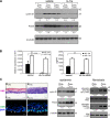
Reduced sIL-1ra in fibroblasts potentiates epidermal keratinocyte proliferation. (A) Immunoblot analysis of epidermis from indicated OTCs treated with either vehicle (PBS) or 50 ng/ml exogenous IL-1ra. Cell proliferation was measured using PCNA and cyclin D1. Values below each band were derived as described in Fig. 3 C. (B) Specific knockdown of sIL-1ra in human fibroblasts. (left) Knockdown efficiency was monitored by qPCR and normalized with control ribosomal protein P0. Specificity of knockdown was assessed by the relative expression level of icIL-1ra. (right) Protein expression of sIL-1ra and icIL-1ra as determined by ELISA. (C) Reduced fibroblasts sIL-1ra increase epidermal proliferation. OTCs were constructed using KCTRL with either control (FCTRL) or sIL-1ra knockdown (FsIL-1ra) fibroblasts. H&E, hematoxylin and eosin staining. (top) Ki67, cell proliferation (white arrows); DAPI, nuclear staining. Bars, 40 µm. (bottom) Bars, 20 µm. (left) Mean numbers of proliferating cells were derived as described in Fig. S1. Broken lines denote epidermal–dermal junction. (right) Immunoblot analysis of keratinocytes and fibroblasts extracted from two independent indicated OTCs. Equal amounts of total protein (50 µg) were resolved, electrotransferred, and probed for the indicated proteins. Values below each band represent the mean fold differences in expression level with respect to KCTRL or FCTRL extracted from KCTRL/FCTRL OTC. **, P < 0.01; ***, P < 0.001. Data are mean ± SD, n = 3.
Figure 6.
Figure 6.
Increased c-Jun binding to AP-1 site of human KGF, GMCSF, and IL-6 gene promoter in FsIL-1ra. ChIP of AP-1–binding site of human KGF (top), GMCSF (middle), and IL-6 (bottom) genes using phospho–c-Jun antibodies. The gene sequence spanning the AP-1 site and a random control sequence were analyzed by PCR in the immunoprecipitated chromatin of FCTRL and FsIL-1ra fibroblasts extracted from K/FCTRL and K/FsIL-1ra OTC, respectively. Preimmune serum was used as a control. qPCR was performed on immunoprecipitates of phospho–c-Jun antibodies and normalized to input (chromatin before immunoprecipitation). Results are represented in fold change as compared with FCTRL extracted from K/FCTRL OTC. M, 100 bp DNA ladder.
Figure 7.
Figure 7.
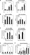
Regulation of IL-1β signaling via PPARβ/δ during wound repair in KO mice. (A and B) Expression level of sIL-1ra (A) and icIL-1ra (B) in WT and KO fibroblasts. WT and KO primary fibroblasts were treated with PPARβ/δ ligand (100 nM GW501516 [GW]) and/or 10 ng/ml IL-1β. The mRNA (left) and protein (right) expression levels were measured by qPCR and ELISA, respectively. (C) Expression level of sIL-1ra in early wound biopsies of KO mice. Wound fluids and biopsies were collected at the indicated day post-wounding (days 1–3, n = 7; day 7, n = 4). Unwounded skin was used as control (ctrl; n = 4). sIL-1ra mRNA and protein levels were determined by qPCR and ELISA, respectively. (D) Expression of sIL-1ra, icIL-1ra, KGF, IL-6, and GMCSF mRNAs from laser capture microdissected (LCM) dermis of WT (n = 3) and KO (n = 4) wound biopsies. Indicated mitogenic factor mRNA levels were analyzed by qPCR and normalized to control ribosomal protein P0. (E) Expression levels of indicated factor mRNAs in vehicle (veh; carboxymethylcellulose)- and IL-1ra–treated (3 × 2.5 µg) wounds of KO and WT mice as compared with corresponding unwounded (control) skin. *, P < 0.05; **, P < 0.01; ***, P < 0.001. Data are mean ± SEM, n = 4.
Figure 8.
Figure 8.
The autonomous and nonautonomous actions of PPARβ/δ. PPARβ/δ confers antiapoptotic properties to keratinocytes and potentiates their migration via AKT1 and Rho GTPases in a cell-autonomous manner (Di Poi et al., 2002; Tan et al., 2007). PPARβ/δ also influences keratinocyte differentiation in a yet unknown mechanism. PPARβ/δ regulates epidermal proliferation in a nonautonomous fashion via a paracrine mechanism. IL-1 constitutively produced by the adjacent keratinocytes activates c-Jun, an obligate partner of AP-1 transcription factor via TAK1, and consequently increases the production of several mitogenic factors. The expression of PPARβ/δ in the underlying fibroblasts attenuates this IL-1 signaling via the production of sIL-1ra. sIL-1ra has little affinity for IL-1R2, which is highly expressed in keratinocytes. Thus, sIL-1ra acts in an autocrine fashion onto the fibroblasts, which expressed the predominant functional IL-1R1. The binding of sIL-1ra to IL-1R1 modulates the IL-1–mediated signaling events and consequently decreases the production of several AP-1–mediated mitogenic factors. The mitogenic factors exert a reduced paracrine effect on the epithelial proliferation via their cognate receptors (R). Therefore, PPARβ/δ in the fibroblasts plays an important homeostatic role in maintaining epidermal homeostasis, the absence of PPARβ/δ resulting in significant epidermal proliferation.

Similar articles

Cited by

References

    1. Arend W.P., Malyak M., Guthridge C.J., Gabay C. 1998. Interleukin-1 receptor antagonist: role in biology.Annu. Rev. Immunol. 16:27–55 - PubMed
    1. Banda N.K., Guthridge C., Sheppard D., Cairns K.S., Muggli M., Bech-Otschir D., Dubiel W., Arend W.P. 2005. Intracellular IL-1 receptor antagonist type 1 inhibits IL-1-induced cytokine production in keratinocytes through binding to the third component of the COP9 signalosome.J. Immunol. 174:3608–3616 - PubMed
    1. Barak Y., Liao D., He W., Ong E.S., Nelson M.C., Olefsky J.M., Boland R., Evans R.M. 2002. Effects of peroxisome proliferator-activated receptor delta on placentation, adiposity, and colorectal cancer.Proc. Natl. Acad. Sci. USA. 99:303–308 - PMC - PubMed
    1. Beanes S.R., Dang C., Soo C., Ting K. 2003. Skin repair and scar formation: the central role of TGF-beta.Expert Rev. Mol. Med. 5:1–22 - PubMed
    1. Burdick A.D., Kim D.J., Peraza M.A., Gonzalez F.J., Peters J.M. 2006. The role of peroxisome proliferator-activated receptor-beta/delta in epithelial cell growth and differentiation.Cell. Signal. 18:9–20 - PubMed

Publication types

MeSH terms