. 2009 Mar 23;184(6):847-62.
doi: 10.1083/jcb.200808124.

N-glycans are direct determinants of CFTR folding and stability in secretory and endocytic membrane traffic

Affiliations

N-glycans are direct determinants of CFTR folding and stability in secretory and endocytic membrane traffic

Rina Glozman et al. J Cell Biol. .

Abstract

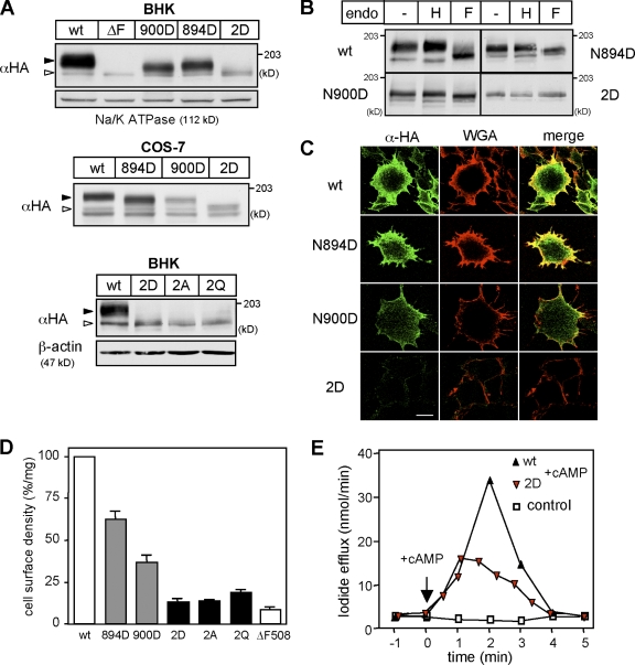
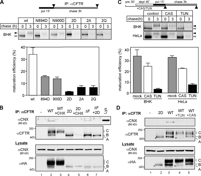
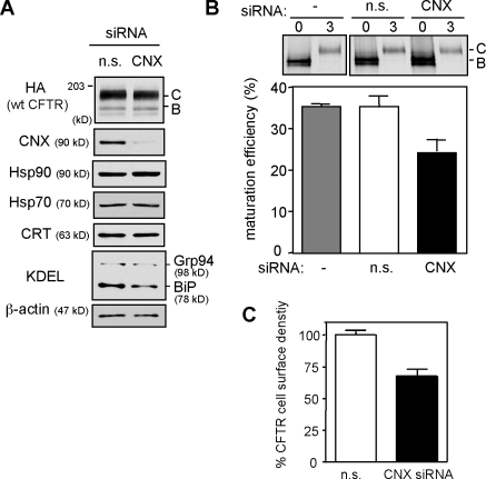
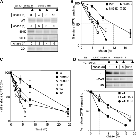
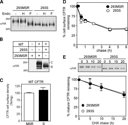
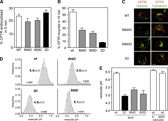
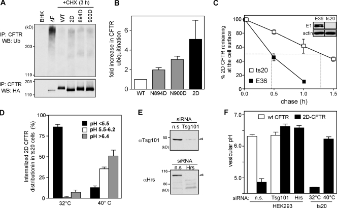
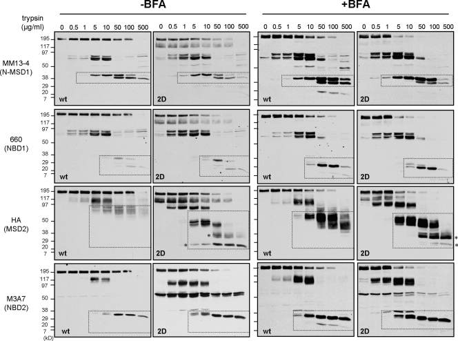
N-glycosylation, a common cotranslational modification, is thought to be critical for plasma membrane expression of glycoproteins by enhancing protein folding, trafficking, and stability through targeting them to the ER folding cycles via lectin-like chaperones. In this study, we show that N-glycans, specifically core glycans, enhance the productive folding and conformational stability of a polytopic membrane protein, the cystic fibrosis transmembrane conductance regulator (CFTR), independently of lectin-like chaperones. Defective N-glycosylation reduces cell surface expression by impairing both early secretory and endocytic traffic of CFTR. Conformational destabilization of the glycan-deficient CFTR induces ubiquitination, leading to rapid elimination from the cell surface. Ubiquitinated CFTR is directed to lysosomal degradation instead of endocytic recycling in early endosomes mediated by ubiquitin-binding endosomal sorting complex required for transport (ESCRT) adaptors Hrs (hepatocyte growth factor-regulated tyrosine kinase substrate) and TSG101. These results suggest that cotranslational N-glycosylation can exert a chaperone-independent profolding change in the energetic of CFTR in vivo as well as outline a paradigm for the peripheral trafficking defect of membrane proteins with impaired glycosylation.


Figures

Figure 1.
Figure 1.
N-glycosylation defect reduces the cell surface expression of CFTR. (A) Expression levels of CFTR-3HA variants in stably (BHK) and transiently (COS-7) transfected cells were visualized by immunoblotting. Na+/K+-ATPase and β-actin were used as a loading control. Open and closed arrowheads indicate core-glycosylated/nonglycosylated CFTR and complex-glycosylated CFTR, respectively. The ΔF508-CFTR mutant remains core glycosylated. (B) Endo H (H) and PNGase F (F) sensitivity of CFTR variants was assessed by immunoblotting with anti-HA Ab after the incubation of BHK cell lysates with or without endo for 3 h at 33°C. (C) Cell surface expression of CFTR-3HA variants in BHK cells was examined by immunostaining with anti-HA Ab without permeabilization. Plasma membrane was stained with Alexa Fluor 594–WGA. (D) Cell surface density of CFTR variants in BHK cells was measured by anti-HA Ab–binding assay and expressed as a percentage of the wt CFTR normalized for cellular proteins. (E) 2D-CFTR is active in BHK cells, as measured by iodide efflux assay. Error bars indicate mean ± SEM. Bar, 10 µm.
Figure 2.
Figure 2.
Defective N-glycosylation severely prevents the productive folding of CFTR. (A) The pulse-labeled CFTR folding efficiency was determined by the production of the complex-glycosylated form or by the remaining amount of protease resistant (2D) after 3-h chase and expressed as a percentage of the initial radioactivity measured by phosphorimage analysis (n = 3–6). (B) CNX binding to CFTR immunoprecipitates was measured by immunoblotting with anti-CNX Ab. Nonnative folding intermediates (immature form) of wt and 2D-CFTR were eliminated with CHX for 3 h. (C) Differential effect of CAS and TUN on wt CFTR folding efficiency measured as in A in BHK and HeLa cells. Cells were treated with 1 mM CAS or 10 µg/ml TUN for 90 min before and during the pulse labeling (n = 3–5). (D) TUN and CAS treatment (4 h) prevents CNX association with nonnative wt CFTR determined as in B. Asterisks indicate the nonglycosylated wt CFTR. A, B, and C indicate nonglycosylated CFTR, core-glycosylated CFTR, and complex-glycosylated CFTR, respectively. Open and closed arrowheads indicate the core-glycosylated/nonglycosylated CFTR and complex-glycosylated CFTR, respectively. IP, immunoprecipitation; pre, preincubation; deple, depletion; pul, pulse label. Error bars indicate mean ± SEM.
Figure 3.
Figure 3.
CNX knockdown partially inhibits CFTR folding. (A) Selective down-regulation of CNX with 100 nM siRNA in CFTR-expressing HeLa cells. Equal amounts of nonspecific (n.s.) and CNX siRNA–treated lysates were analyzed by immunoblotting. C and B indicate complex-glycosylated CFTR and core-glycosylated CFTR, respectively. (B) Folding yield of wt CFTR in CNX-depleted HeLa cells was measured by pulse-chase experiments (n = 3). (C) Effect of CNX knockdown on the cell surface density of CFTR in HeLa cells (n = 3). Error bars indicate mean ± SEM.
Figure 4.
Figure 4.
N-glycans are required for CFTR stability in post-Golgi compartments. (A and B) Turnover of mature CFTR variants was measured after 40-min pulse labeling (pul) and 3-h chase by immunoprecipitation and phosphorimage analysis (n = 5). (C) The cell surface stability of CFTR-3HA variants was determined by anti-HA Ab–binding assay. The disappearance of anti-HA Ab bound to cell surface CFTR was monitored at 37°C (n = 4). (D) The effect of TUN and CAS on the turnover of mature wt CFTR in post-Golgi compartments (n = 3). Glycosylation inhibitors were present during the preincubation (90 min) and the radioactive pulse (40 min) as indicated. Dashed lines in B and D indicate the half-life of mature CFTR. Arrowheads in the scheme in A and D indicate the time when the sample was collected. Error bars indicate mean ± SEM.
Figure 5.
Figure 5.
Core glycosylation is sufficient for the folding and stability of CFTR. (A) Endo H (H) and PNGase F (F) sensitivity of wt CFTR in HEK293S cells was assessed by immunoblotting with anti-HA Ab after the incubation of cell lysates for 3 h at 33°C. HEK293MSR cells were used as control. Steady-state level (B) and cell surface density (C) of wt CFTR in HEK293S cells (n = 3). (D) Cell surface stability of wt CFTR in HEK293S cells was measured by anti-HA–binding assay (n = 3). MSR and S indicate HEK293MSR and HEK293S cells, respectively. C and B indicate complex-glycosylated CFTR and core-glycosylated CFTR, respectively. (E) Turnover rates of the mature complex-glycosylated CFTR and core-glycosylated CFTR in HEK293MSR and HEK293S cells, respectively. Metabolic turnover of CFTR was measured by CHX chase after an initial CHX treatment (3 h) to eliminate the ER resident folding intermediates (n = 3). Error bars indicate mean ± SEM.
Figure 6.
Figure 6.
Lysosomal targeting and inefficient endocytic recycling account for the cell surface instability of glycosylation-deficient CFTR. (A) Internalization rate of CFTR variants in BHK cells was measured by anti-HA Ab uptake assay as described in Materials and methods (n = 3–6). (B) Endocytic recycling of CFTR variants in BHK cells was measured by the biotin-NeutrAvidin sandwich assay as described in Materials and methods (n = 3). (C) Colocalization of internalized CFTR with FITC-dextran–loaded lysosomes and FITC-transferrin–labeled early endosomes was analyzed by laser confocal microscopy as described in Materials and methods. CFTR variants were internalized with anti-HA Ab complexed to TRITC-conjugated anti–mouse IgG or Fab for 1 h at 37°C and chased in Ab-free medium for 30 min. Single optical sections obtained by laser confocal microscopy were taken, and only overlay images are shown. (D) The pH of the endocytic vesicles containing CFTR variants was measured by single-cell fluorescence ratio image analysis using in situ calibration curve. The pH of >300 vesicles was determined in each experiment as described in Materials and methods. (E) The mean pH of vesicular populations containing internalized CFTR variants in BHK, HEK293MSR (MSR), and HEK293S (S) cells from 3–4 independent experiments. Error bars indicate mean ± SEM. Bar, 10 µm.
Figure 7.
Figure 7.
Ubiquitination acts as sorting signal directing glycosylation-deficient CFTR into lysosome. (A) Ubiquitination level of CFTR variants in post-Golgi compartments of BHK cells was measured by denaturing immunoprecipitation (IP) and immunoblotting with anti-Ub Ab. Five- to sixfold more cells were used for the glycosylation-deficient CFTR isolation. Protein loading was adjusted to have a comparable amount of wt and glycosylation-deficient CFTR. (B) Relative ubiquitination was measured by densitometry after normalization for the amount of precipitated CFTR. (C) Cell surface stability of 2D-CFTR in ts20 and E36 cells was measured by anti-HA Ab binding. Cells were exposed to 40°C for 2.5 h to inactive E1, as verified by immunoblotting (inset). Dashed lines in the plot indicate the half-life of cell surface CFTR. (D) The pH of the endocytic vesicles containing 2D-CFTR in ts20 cells preincubated at 32 or 40°C for 2.5 h was measured at 37°C by FRIA. The percentage of 2D-CFTR residing in vesicles with the indicated pH is plotted. (E) Down-regulation of Hrs and TSG101 by siRNA was verified in HEK cells by immunoblotting. Equal amounts of proteins were loaded. (F) The mean pH of vesicles containing wt or 2D-CFTR variants in HEK cells transfected with 50 nM TSG101, Hrs, or nonspecific (n.s.) siRNA as described in Materials and methods. Sorting of 2D-CFTR in ts20 cells preincubated at 32 or 40°C for 2.5 h was measured by FRIA (n = 3). WB, Western blot. Error bars indicate mean ± SEM.
Figure 8.
Figure 8.
N-glycosylation increases the protease resistance of the mature CFTR. Limited trypsinolysis of the wt and 2D-CFTR. Isolated microsomes from BHK cells treated with or without 5 µg/ml BFA for 24 h were exposed to trypsin. Proteolytic patterns were visualized by immunoblotting with domain-specific (shown in parentheses) anti-CFTR antibodies. Dashed regions designate putative fragments containing the Ab-specific domains validated previously (Du et al., 2005; Cui et al., 2007). The glycosylation defect provoked increased protease susceptibility of the MSD2 relative to wt in the absence as well as in the presence of BFA. The asterisks designate novel MSD2 degradation intermediate in the 2D-CFTR.
Figure 9.
Figure 9.
N-glycosylation enhances the thermal stability of CFTR. (A and B) Determination of the mature wt and 2D-CFTR thermoaggregation propensity. wt (top) and 2D-CFTR (bottom) were solubilized in RIPA buffer after CHX treatment of the cells for 3 h. Cell lysates were mixed with LSB and incubated at the indicated temperature for 5 min. Aggregates were pelleted by centrifugation, and soluble fractions were analyzed with immunoblotting. Dashed regions indicate the monomeric mature form of wt and 2D-CFTR. (B) The monomeric wt and 2D-CFTR were quantified with densitometry on immunoblots shown in A and expressed as the percentage of the amount detected at 37°C incubation (n = 3). (C and D) The effect of low temperature (26°C for 24 h) incubation on the steady-state level (C) and cell surface density (D) of 2D-CFTR, as measured by Western blotting and anti-HA Ab binding, respectively (n = 3). Open and closed arrowheads indicate the core-glycosylated/nonglycosylated and complex-glycosylated CFTR, respectively. Error bars indicate mean ± SEM.

Similar articles

Cited by

References

    1. Agashe V.R., Guha S., Chang H.C., Genevaux P., Hayer-Hartl M., Stemp M., Georgopoulos C., Hartl F.U., Barral J.M. 2004. Function of trigger factor and DnaK in multidomain protein folding: increase in yield at the expense of folding speed.Cell. 117:199–209 - PubMed
    1. Arvan P., Zhao X., Ramos-Castaneda J., Chang A. 2002. Secretory pathway quality control operating in Golgi, plasmalemmal, and endosomal systems.Traffic. 3:771–780 - PubMed
    1. Asano T., Takata K., Katagiri H., Ishihara H., Inukai K., Anai M., Hirano H., Yazaki Y., Oka Y. 1993. The role of N-glycosylation in the targeting and stability of GLUT1 glucose transporter.FEBS Lett. 324:258–261 - PubMed
    1. Balch W.E., Morimoto R.I., Dillin A., Kelly J.W. 2008. Adapting proteostasis for disease intervention.Science. 319:916–919 - PubMed
    1. Barriere H., Nemes C., Du K., Lukacs G.L. 2007. Plasticity of polyubiquitin recognition as lysosomal targeting signals by the endosomal sorting machinery.Mol. Biol. Cell. 18:3952–3965 - PMC - PubMed

Publication types

MeSH terms

Substances

LinkOut - more resources