. 2009 Mar 23;184(6):881-94.
doi: 10.1083/jcb.200808041.

A role for ubiquitin ligases and Spartin/SPG20 in lipid droplet turnover

Affiliations

A role for ubiquitin ligases and Spartin/SPG20 in lipid droplet turnover

Scott W Eastman et al. J Cell Biol. .

Abstract

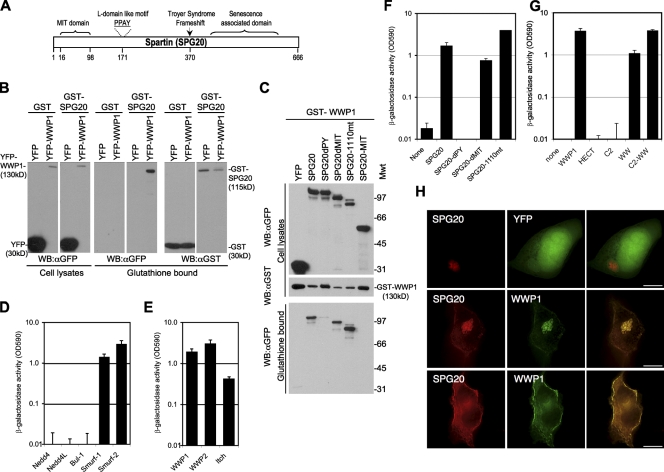
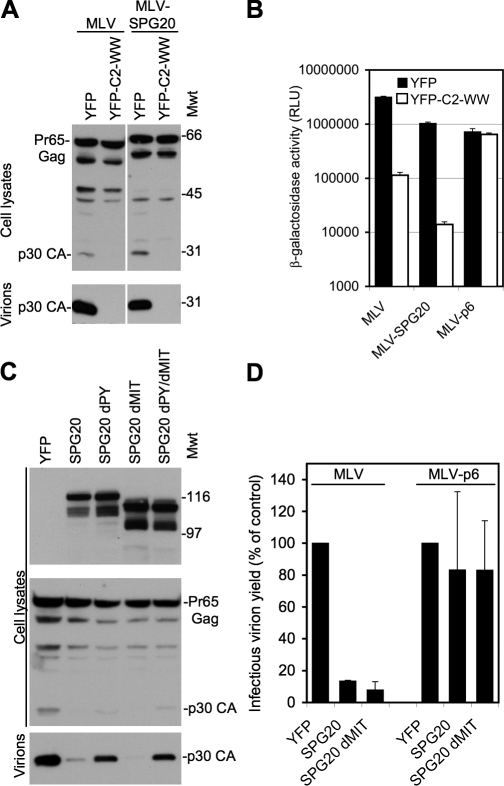
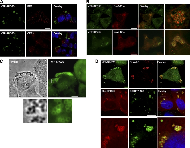
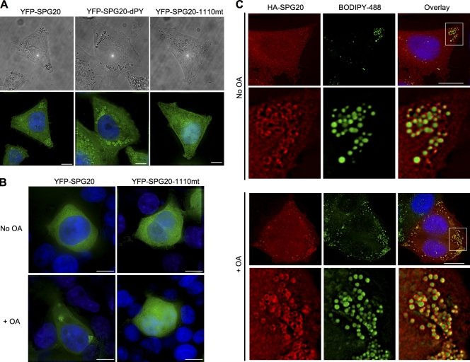
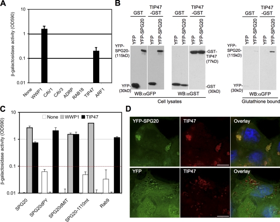
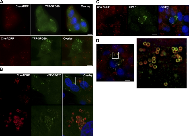
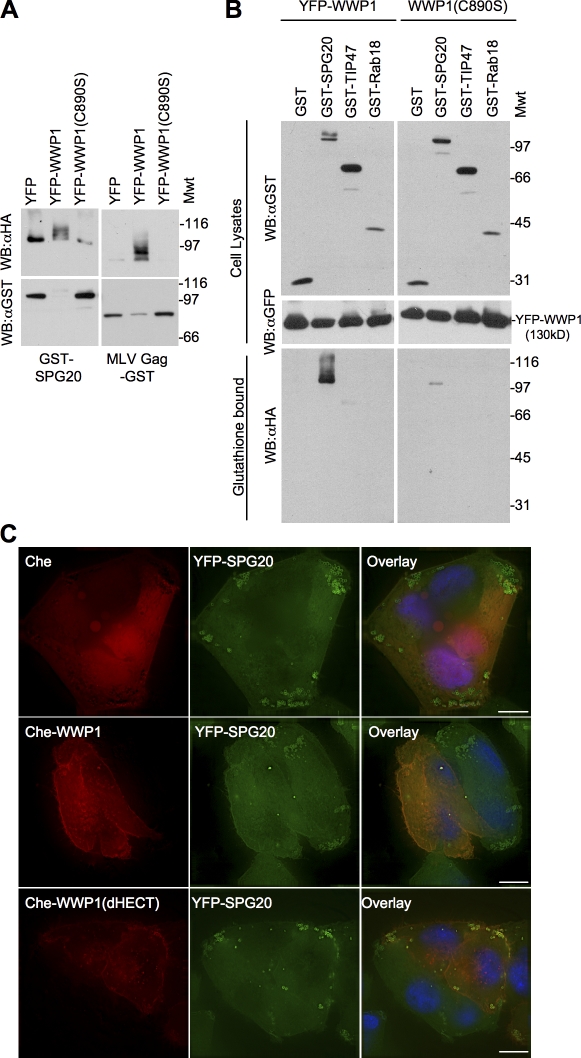
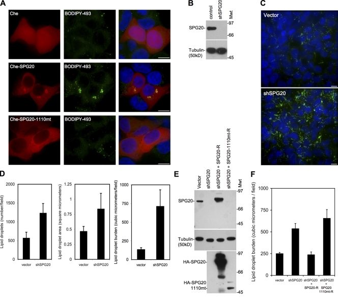
HECT (homologous to the E6AP C terminus) ubiquitin ligases have diverse functions in eukaryotic cells. In screens for proteins that bind to the HECT ubiquitin ligase WWP1, we identified Spartin, which is also known as SPG20. This protein is truncated in a neurological disease, Troyer syndrome. In this study, we show that SPG20 associates with the surface of lipid droplets (LDs) and can regulate their size and number. SPG20 binds to another LD protein, TIP47, and both proteins compete with an additional LD protein, adipophilin/adipocyte differentiation-related protein, for occupancy of LDs. The mutant SPG20 present in Troyer syndrome does not possess these activities. Depletion of SPG20 using RNA interference increases the number and size of LDs when cells are fed with oleic acid. Binding of WWP1 to SPG20 and the consequent ubiquitin transfer remove SPG20 from LDs and reduce the levels of coexpressed SPG20. These experiments suggest functions for ubiquitin ligases and SPG20 in the regulation of LD turnover and potential pathological mechanisms in Troyer syndrome.


Figures

Figure 1.
Figure 1.
SPG20 interacts with the WW domains of WWP1 via its PPxY motif. (A) Schematic representation of the Spartin/SPG20 protein showing the MIT domain (aa 16–98), PPAY sequence (aa 171–174), and Troyer syndrome frameshift mutant (deletion of nt 1,110 at aa 370 to truncate the 666-aa protein at aa 398). (B) Coprecipitation of WWP1 and SPG20. Unfused GST or GST-SPG20 was coexpressed with YFP or YFP-WWP1 in 293T cells, and cell lysates as well as the glutathione-bound fraction were analyzed by Western blotting with α-GST and α-GFP antibodies. (C) GST-WWP1 coprecipitation experiments with YFP fused to wild-type SPG20, SPG20 lacking the PPxY domain (SPG20dPY), SPG20 lacking the MIT domain (SPG20dMIT), the Troyer syndrome mutant SPG20 (SPG20-1110mt), or the isolated SPG20 MIT domain (SPG20-MIT). Mwt, molecular weight; WB, Western blot. (D–G) Yeast two-hybrid analysis of interactions between SPG20 and HECT ubiquitin ligases. β-Galactosidase activity measured in yeast transformed with Gal4 DNA-binding domain fusion proteins and VP16 activation domain fusion proteins expressed in optical density units (OD590). (D) Yeast two-hybrid analysis interactions between a Gal4-SPG20 and WW domains of Nedd4, Nedd4-L, Bul-1, Smurf1, and Smurf2. (E) Yeast two-hybrid analysis interactions between a Gal4-SPG20 and full-length WWP1, WWP2, and Itch. (F) Yeast two-hybrid analysis of Gal4-WWP1 binding to the various SPG20 mutants described in C. (G) Yeast two-hybrid analysis of Gal4-SPG20 binding to the various domains of WWP1: the N-terminal C2 domain, the central (PPxY binding) WW domains, and the C-terminal catalytic HECT domain. (H) Images of 293T cells transiently overexpressing CFP-SPG20 and either unfused YFP (top) or YFP-WWP1 (middle and bottom). Two different optical sections from a stack of deconvolved images are shown in the middle and bottom panels. Error bars represent the standard deviation of the mean. Bars, 10 µm.
Figure 2.
Figure 2.
Virus particle release assays to demonstrate interactions between SPG20 and HECT ubiquitin ligases within cells. (A) Release of virions from cells transfected with MLV constructs encoding either the unmodified PPPY domain or one in which the PPPY motif was replaced with a region encompassing the SPG20-PPxY sequence (MLV-SPG20). Virions were generated in the presence or absence of the WWP1dHECT protein, and blots were probed with an α-MLV capsid antibody. Mwt, molecular weight. (B) Measurement of infectious virions generated by MLV, MLV-SPG20, or an MLV encoding the HIV-p6–derived PTAP motif (MLV-p6). Virions containing the pMSCV/Tat vector were generated in the presence or absence of the WWP1dHECT protein and were quantitated by measuring β-galactosidase activity after inoculation of TZM cells. RLU, relative light unit. (C) Western blot analysis of cell lysates and released MLV virion particles from 293T cells cotransfected with an unmodified MLV provirus along with YFP alone or YFP fused to SPG20, SPG20dPY, SPG20dMIT, or SPG20dPYdMIT. Blots were probed with α-GFP (top) or α-MLV capsid (bottom) antibodies. (D) Infectious virion release from cells transfected with MLV or MLV-p6 in the presence or absence of SPG20. Infectious virions were measured using TZM indicator cells as in B and are expressed as a percentage of control uninhibited virion release. Error bars represent the standard deviation of the mean.
Figure 3.
Figure 3.
SPG20 localizes to LDs. (A) Immunofluorescent localization of endosome markers (EEA1 and CD63; red) in a cell line (HeLa/YFP-SPG20) stably expressing a YFP-SPG20 fusion protein (green; images represent projections of deconvolved image stacks). (B) Localization of Che-tagged caveolin proteins (Cav1-Che and Cav3-Che; red) after transient expression in HeLa/YFP-SPG20 cells. Expanded views of the boxed areas in the overlay images are shown to the right. (C) Phase-contrast (left) and fluorescent (right) images of HeLa/YFP-SPG20 cells. An expanded view of the boxed area is shown below. (D) Staining of HeLa cells with LD markers. HeLa/YFP-SGP20 cells were stained with the LD marker Oil red O (top), whereas HeLa/Che-SPG20 cells were stained using the LD marker BODIPY 493/503 (middle). An expanded view of the boxed area in the BODIPY 493/503–stained HeLa/Che-SPG20 is shown in the bottom panels. Bars, 10 µm.
Figure 4.
Figure 4.
The C-terminal region of SPG20 that is absent in the Troyer syndrome variant is required for LD targeting. (A) HeLa cell lines stably expressing YFP-SPG20, YFP-SPG20dPY, or YFP-SPG20-1110mt were visualized using phase contrast (top) and fluorescence (bottom). LDs, which are present in all cells, can be seen as dark spheres and are more prominent in the presence of YFP-SPG20 and YFP-SPG20dPY. (B) 293T cells stably expressing YFP-SPG20 (left) or YFP-SGP20-1110mt (right) under normal culture conditions (top) or after incubation for 18 h with OA (bottom). Note that in 293T cells, expression of the YFP fusion proteins is lower than in HeLa cells, and YFP-SPG20 appears diffuse under normal culture conditions (top left). (C) Localization of HA-tagged SPG20. HeLa cells stably expressing HA-SPG20 were cultured in the absence (top) or presence (bottom) of OA and stained with antibody to the HA epitope tag (red) and with BODIPY 493/53 (green). An expanded view of LDs present in the area of the images bounded by the white boxes is shown below each image set. Bars, 10 µm.
Figure 5.
Figure 5.
SPG20 interacts with TIP47. (A) Directed yeast two-hybrid analysis of interactions between Gal4-SPG20 and candidate LD-associated proteins (Cav1, Cav3, ADRP, Rab18, TIP47, and Arf-1) fused to the VP16 activation domain. WWP1 is included as a positive control. (B) GST coprecipitation of YFP-SPG20 and TIP47-GST. Unfractionated 293T cell lysates and proteins bound to glutathione-agarose were detected by Western blotting with α-GFP and α-GST antibodies. WB, Western blot. (C) Yeast two-hybrid analysis of interactions between SPG20 and TIP47. The DNA-binding constructs containing full-length SPG20, SPG20dPY, SPG20dMIT, and SPG20-1110mt were tested against the VP16 activation domain fused to WWP1 or TIP47. Gal4-Rab9 was included in the experiment as a control for TIP47 binding. (D) Colocalization of SPG20 and TIP47. Endogenous TIP47 is stained with an α-TIP47 monoclonal antibody in HeLa cells stably expressing either YFP-SPG20 (top) or unfused YFP (bottom). Error bars represent the standard deviation of the mean. Bars, 10 µm.
Figure 6.
Figure 6.
Reciprocal exclusion of SPG20, TIP47, and ADRP from LDs. (A) Expression of ADRP displaces SPG20 from LDs and vice versa. HeLa/YFP-SPG20 cells were transduced with a retroviral vector expressing Che-ADRP and visualized several days after infection. The images shown represent a projection of a deconvolved vertical stack of images, and a field containing both Che-ADRP–positive and Che-ADRP–negative cells is shown (top). The reciprocal experiment, in which a HeLa Che-ADRP cell line was transduced with a YFP-SPG20–expressing retroviral vector, is shown in the bottom panels. (B) Coresidence of Che-ADRP and YFP-SPG20 in cells expressing lower levels of YFP-SPG20. An expanded view of the boxed area is shown below. (C and D) HeLa/Che-ADRP cells stained with antibodies against endogenous TIP47 (green). Note that in C, TIP47 localization is only clearly visible in cells expressing no or low levels of Che-ADRP. In D, an expanded area of a cell (boxed area) expressing moderate levels of ADRP is shown, revealing coexistence of TIP47 and ADRP on individual LDs but apparently occupying distinct areas of the LD surface. Bars, 10 µm.
Figure 7.
Figure 7.
WWP1 can ubiquitinate SPG20 and prevent its accumulation on LDs. (A) Ubiquitin transfer experiment performed in 293T cells cotransfected with GST-SPG20 or MLV Gag–GST along with HA-ubiquitin and unfused YFP, YFP-WWP1, or catalytically inactive YFP-WWP1 (C890S). Glutathione-bound proteins were analyzed by Western blotting with α-HA and α-GST antibodies. Mwt, molecular weight. WB, Western blot. (B) Similar to A except that cell lysates and glutathione-bound fractions were analyzed by Western blotting. 293T cells transfected with GST-SPG20, GST-TIP47, or GST-Rab18 along with YFP-WWP1 or YFP-WWP1 (C890S) as well as HA-ubiquitin. GST-bound proteins were probed with antibodies against the HA tag (bottom) to detect specific ubiquitin transfer. Cellular lysates were probed with antibodies against GST (top) and YFP (middle) to detect the corresponding fusion proteins. (C) Overexpression of WWP1 depletes SPG20 from LDs. HeLa YFP-SPG20 cells were transduced with retroviruses expressing unfused Che (top), Che-WWP1 (middle), or Che-WWP1dHECT (bottom). Bars, 10 µm.
Figure 8.
Figure 8.
Over- or underexpression of SPG20 perturbs LDs. (A) 293T cells transfected with Che (top), Che-SPG20 (middle), or Che-SPG20-1110mt (bottom) and stained with the LD marker BODIPY 493/503. (B) SPG20-depleted cells generated by transduction of 293T cells with lentiviral vectors expressing an shRNA targeting SPG20 (shSPG0). Western blot analysis of cell lysates probed with an antibody against SPG20 (top) and against tubulin (bottom) as a loading control. Mwt, molecular weight. (C) Representative fields of control vector–containing (top) and shSPG20-containing (bottom) 293T cells cultured in the presence of 500 µM OA for 48 h and stained with BODIPY 493/503 to detect LDs (green). (D) The total number and size of LDs were quantified in 10 randomly selected fields using the softWoRx software. (E and F) Similar experiments as shown in B–D elaborated by the inclusion of shSPG20 cells reconstituted with an shRNA-resistant form of wild-type HA-tagged SPG20 (SPG20-R) or the Troyer syndrome variant (SPG20-1110mt-R). (E) Western blots probed with antibodies to SPG20 (top), tubulin (loading control; middle), and HA-tagged proteins (bottom). (F) LD burden in SPG20-depleted/reconstituted cells determined as in D. Error bars represent the standard deviation of the mean. Bars, 10 µm.

Similar articles

Cited by

References

    1. Agarwal A.K., Garg A. 2004. Seipin: a mysterious protein.Trends Mol. Med. 10:440–444 - PubMed
    1. Bakowska J.C., Jenkins R., Pendleton J., Blackstone C. 2005. The Troyer syndrome (SPG20) protein spartin interacts with Eps15.Biochem. Biophys. Res. Commun. 334:1042–1048 - PubMed
    1. Bakowska J.C., Jupille H., Fatheddin P., Puertollano R., Blackstone C. 2007. Troyer syndrome protein spartin is mono-ubiquitinated and functions in EGF receptor trafficking.Mol. Biol. Cell. 18:1683–1692 - PMC - PubMed
    1. Bakowska J.C., Wang H., Xin B., Sumner C.J., Blackstone C. 2008. Lack of spartin protein in Troyer syndrome: a loss-of-function disease mechanism? Arch. Neurol. 65:520–524 - PMC - PubMed
    1. Bartz R., Zehmer J.K., Zhu M., Chen Y., Serrero G., Zhao Y., Liu P. 2007. Dynamic activity of lipid droplets: protein phosphorylation and GTP-mediated protein translocation.J. Proteome Res. 6:3256–3265 - PubMed

Publication types

MeSH terms