. 2009 Mar 23;184(6):923-33.
doi: 10.1083/jcb.200805063.

Akt regulates L-type Ca2+ channel activity by modulating Cavalpha1 protein stability

Affiliations

Akt regulates L-type Ca2+ channel activity by modulating Cavalpha1 protein stability

Daniele Catalucci et al. J Cell Biol. .

Erratum in

  • J Cell Biol. 2013 Mar 18;200(6):851

Abstract

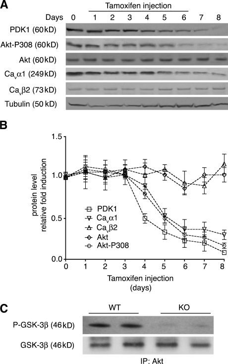
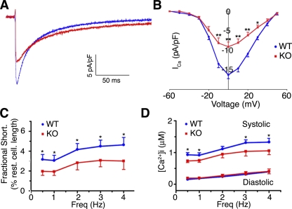
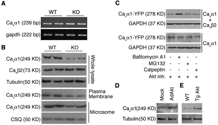
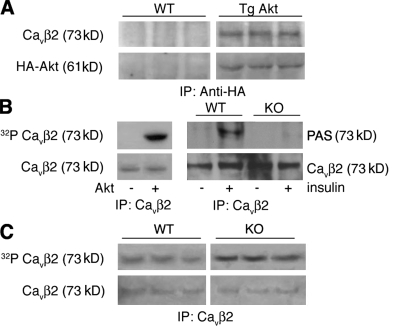
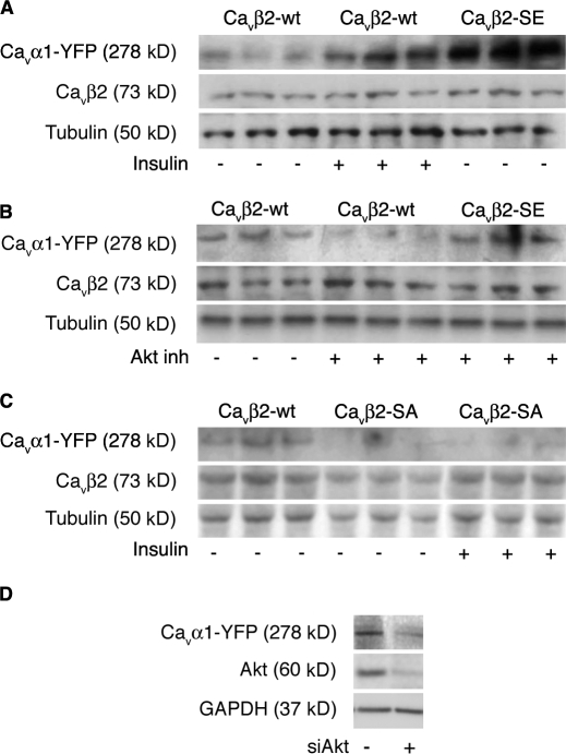
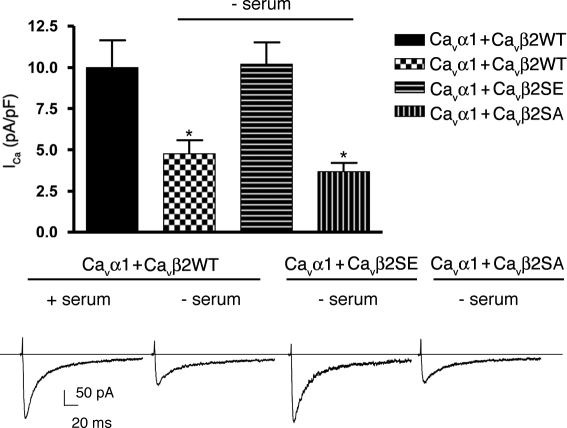
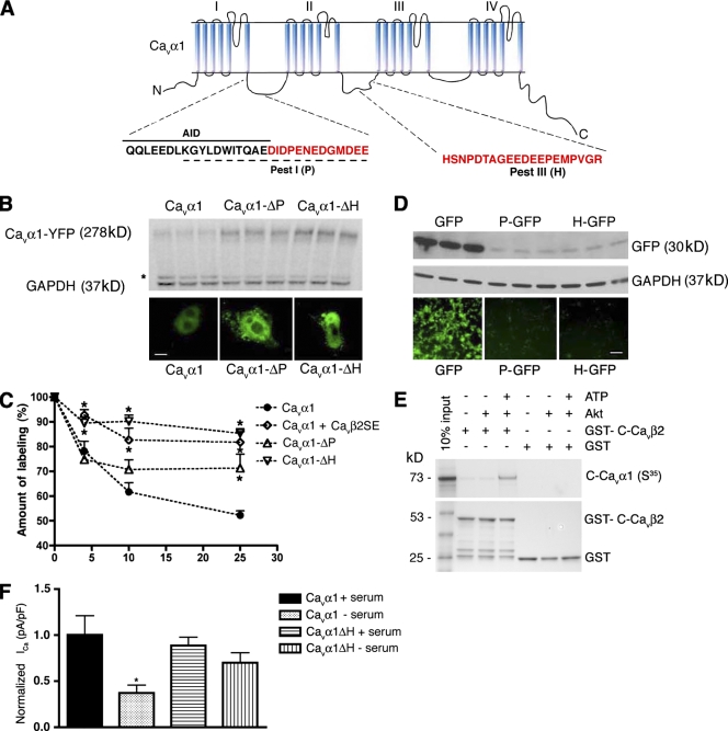
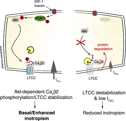
The insulin IGF-1-PI3K-Akt signaling pathway has been suggested to improve cardiac inotropism and increase Ca(2+) handling through the effects of the protein kinase Akt. However, the underlying molecular mechanisms remain largely unknown. In this study, we provide evidence for an unanticipated regulatory function of Akt controlling L-type Ca(2+) channel (LTCC) protein density. The pore-forming channel subunit Ca(v)alpha1 contains highly conserved PEST sequences (signals for rapid protein degradation), and in-frame deletion of these PEST sequences results in increased Ca(v)alpha1 protein levels. Our findings show that Akt-dependent phosphorylation of Ca(v)beta2, the LTCC chaperone for Ca(v)alpha1, antagonizes Ca(v)alpha1 protein degradation by preventing Ca(v)alpha1 PEST sequence recognition, leading to increased LTCC density and the consequent modulation of Ca(2+) channel function. This novel mechanism by which Akt modulates LTCC stability could profoundly influence cardiac myocyte Ca(2+) entry, Ca(2+) handling, and contractility.


Figures

Figure 1.
Figure 1.
Alteration of Ca2+ handling proteins in PDK1 KO cardiomyocytes. (A and B) Western blot (A) and densitometric (B) analyses of ventricular homogenates along a time course of tamoxifen inductions (day 1–6 treatment is indicated by the line in A) using various antibodies. A representative experiment is shown (n = 3). Error bars show SD. (C) Total Akt activity in WT and KO cardiomyocyte lysates assayed using a GSK-3β/α Akt-specific substrate. IP, immunoprecipitation.
Figure 2.
Figure 2.
Impaired intracellular Ca2+ handling and contractility in PDK1 KO cardiomyocytes. (A and B) Smaller Ca2+ current in KO cardiomyocytes. (A) Whole cell representative ICa,L currents normalized for difference in cell size. (B) ICa,L I-V current/voltage relationships (n = 12; *, P < 0.05; **, P < 0.01). (C and D) Cardiomyocyte contraction and Ca2+ transients at different stimulation frequencies. (C) Cardiomyocyte shortening is decreased in KO compared with WT cardiomyocytes (*, P < 0.05; ANOVA). (D) Ca2+ frequency relationship indicates smaller peak systolic but not diastolic Ca2+ in KO compared with WT cells (*, P < 0.05; ANOVA). Error bars show SEM.
Figure 3.
Figure 3.
Akt mediates regulation of Cavα1 protein density at the plasma membrane. (A) RT-PCR analysis of Cavα1 mRNA expression from WT and KO ventricular extracts. GAPDH served as a loading control. (B) Western blot analysis of whole lysate, membrane, and microsomal fractions from WT and KO ventricular extracts. CSQ, calsequestrin. (C) YFP-Cavα1–transfected COS-7 cells alone or in combination with Cavβ2 expression vector were serum starved and treated with Akt inhibitor (Akt inh.) and 1 µM bafilomycin-A1, 25 µM MG132, or 25 µM calpeptin. 6 h after drug administration, cell lysates were prepared and subjected to Western blot analysis for YFP. GAPDH served as a loading control. (D and E) Cavα1 protein levels in KO cardiomyocytes infected with empty (mock) or active E40K-Akt (AdAkt)–expressing adenoviral vector (D) and in whole lysates of WT and E40K-Akt (Tg Akt) hearts (E). Representative experiments are shown (n = 4).
Figure 4.
Figure 4.
Akt interacts with and phosphorylates Cavβ2. (A) Coimmunoprecipitation assay of Akt and Cavβ2. Ventricular homogenates from WT and HA–E40K-Akt Tg mice (Tg Akt) immunoprecipitated with antibodies against HA and immunoblotted for Cavβ2 as well as HA as a control. (B) Examination of Cavβ2 phosphorylation by Akt. In vitro kinase assays were performed with immunoprecipitated Cavβ2 incubated with recombinant active Akt and 32P-labeled ATP (left) or immunoprecipitated Cavβ2 from WT and KO cardiac extracts from mice treated or not treated with 1 mU/g insulin using phospho-Akt substrate (PAS) antibody (right). (C) Back phosphorylation assay of Cavβ2 from WT and KO hearts. Immunoprecipitated Cavβ2 from solubilized membranes was in vitro back phosphorylated using recombinant active Akt and [γ32]ATP. Precipitate amounts were assayed for [32P]Cavβ2 and total Cavβ2. Representative experiments are shown (n = 4). IP, immunoprecipitation.
Figure 5.
Figure 5.
Akt phosphorylation of Cavβ2 protects Cavα1 from protein degradation. (A–C) YFP-Cavα1–cotransfected 293T cells with the indicated mutant variant of Cavβ2. Cells were serum starved overnight and treated with 100 µM insulin (A and C) or 5 µM Akt inhibitor (Akt inh; B) as indicated. The expression of YFP-Cavα1 in lysates was monitored by Western blot analysis with anti-YFP antibody and normalized based on transfection efficiency (Cavβ2) and protein amount (tubulin; n = 3). (D) Cavα1- and Cavβ2-cotransfected 293T cells were treated with siAkt-expressing vector as indicated. 3 d after transfection, cell lysate was tested by Western blot analysis. Protein loading was normalized to GAPDH levels. Representative experiments are shown (n = 3).
Figure 6.
Figure 6.
Akt phosphorylation of Cavβ2 preserves Cavα1 currents. Ca2+ currents recorded in cotransfected tsA-201 cells with YFP-Cavα1 and Cavβ2-WT, Cavβ2-SE, or Cavβ2-SA mutant cultivated for 36 h in the presence or absence of 10% fetal bovine serum. Currents were recorded 1–2 min after the whole cell configuration was achieved (i.e., after stabilization of the current) and were elicited by a 0-mV depolarization of 200-ms duration applied from a holding potential of −80 mV. Currents are normalized to cell capacitance (current density, picoampere/picofarad). Representative current traces are shown. n > 35 at each condition (*, P < 0.05 compared with YFP-Cavα1; ANOVA). Error bars show SEM.
Figure 7.
Figure 7.
Rapid protein degradation PEST sequences determine Cavα1 protein instability. (A) Schematic representation of Cavα1 mapping the AID and PEST sequences in the I–II and II–III cytosolic loops. Deleted PEST sequences (P and H) are highlighted in red. (B) Western blot and immunofluorescence analyses showing relative levels of WT and PEST-deleted mutants of YFP-Cavα1 (n = 3). The asterisk indicates a YFP-Cavα1 degradation fragment. (C) Half-lives of WT Cavα1 subunit (alone or cotransfected with Cavβ2-SE) and its in-frame ΔPEST mutants (Cavα1-ΔP and Cavα1-ΔH) were determined in COS-7 cells. After overnight starvation, transfected cells were pulse chased and analyzed along a time course (*, P < 0.001 compared with Cavα1; ANOVA; n = 3). Error bars show SD. (D) Western blot and immunofluorescence analyses showing relative levels of WT GFP and N-terminal fusion PEST mutants (n = 3). (E) The Cavα1 C terminus interacts with the Akt-phosphorylated GST-Cavβ2 coiled-coil region. Bacterially expressed GST or GST-C-Cavβ2 (Cavβ2, amino acids 480–655) fusion protein and glutathione–Sepharose beads were incubated with equal amounts of in vitro–translated [35S]Met-labeled C-Cavα1 (Cavα1, amino acids 1,477–2,169). Binding occurred only with Akt-phosphorylated GST-C-Cavβ2. Bound proteins were resolved by SDS-PAGE (4–12%). 10% of the input protein in each binding reaction is shown. Coomassie staining of SDS-PAGE is shown in the bottom panel. (F) Ca2+ currents recorded in tsA-201 cells cotransfected with Cavβ2-WT and either Cavα1-WT or Cavα1-ΔH and cultivated for 36 h in the presence or absence of 10% fetal bovine serum. Current densities (picoampere/picofarad) are normalized to the control condition. n > 35 at each condition (*, P < 0.05 compared with Cavα1; ANOVA). Error bars show SEM. Bars: (B) 5 µm; (D) 20 µm.
Figure 8.
Figure 8.
Proposed mechanism. Akt, followed by PDK1 activation, phosphorylates Cavβ2 at the C-terminal coiled-coil domain. The phosphorylation allows association of the C-terminal portion of Cavβ2 with the Cavα1 C-terminal domain. In turn, a conformation shift prevents PEST sequence recognition, stabilizing Cavα1 protein levels. The blue and red ribbons in Cavα1 represent AID and PEST sequences, respectively.

References

    1. Aimond F., Kwak S.P., Rhodes K.J., Nerbonne J.M. 2005. Accessory Kvbeta1 subunits differentially modulate the functional expression of voltage-gated K+ channels in mouse ventricular myocytes. Circ. Res. 96:451–458 - PubMed
    1. Bassani J.W., Bassani R.A., Bers D.M. 1994. Relaxation in rabbit and rat cardiac cells: species-dependent differences in cellular mechanisms. J. Physiol. 476:279–293 - PMC - PubMed
    1. Bayascas J.R., Wullschleger S., Sakamoto K., Garcia-Martinez J.M., Clacher C., Komander D., van Aalten D.M., Boini K.M., Lang F., Lipina C., et al. 2008. Mutation of PDK1 PH domain inhibits PKB/Akt leading to small size and insulin-resistance. Mol. Cell. Biol. 28:3258–3272 - PMC - PubMed
    1. Belles B., Hescheler J., Trautwein W., Blomgren K., Karlsson J.O. 1988. A possible physiological role of the Ca-dependent protease calpain and its inhibitor calpastatin on the Ca current in guinea pig myocytes. Pflugers Arch. 412:554–556 - PubMed
    1. Bers D.M. 2002. Cardiac excitation-contraction coupling. Nature. 415:198–205 - PubMed

Publication types

MeSH terms

Substances