. 2009 Dec;41(6):680-7.
doi: 10.1165/rcmb.2007-0386OC. Epub 2009 Mar 23.

Absence of proteinase-activated receptor-1 signaling in mice confers protection from fMLP-induced goblet cell metaplasia

Affiliations

Absence of proteinase-activated receptor-1 signaling in mice confers protection from fMLP-induced goblet cell metaplasia

Luigi Atzori et al. Am J Respir Cell Mol Biol. 2009 Dec.

Abstract

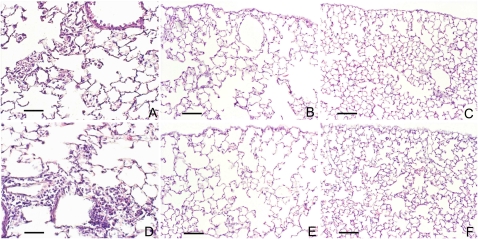
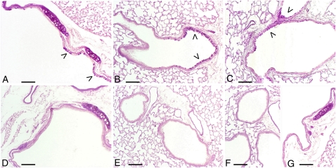
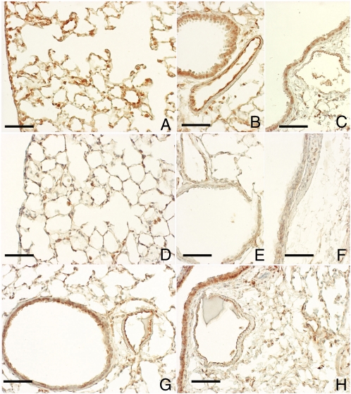
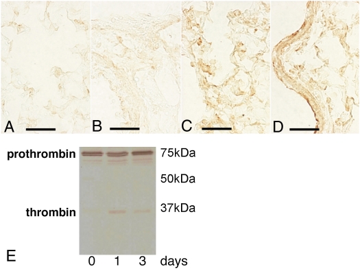
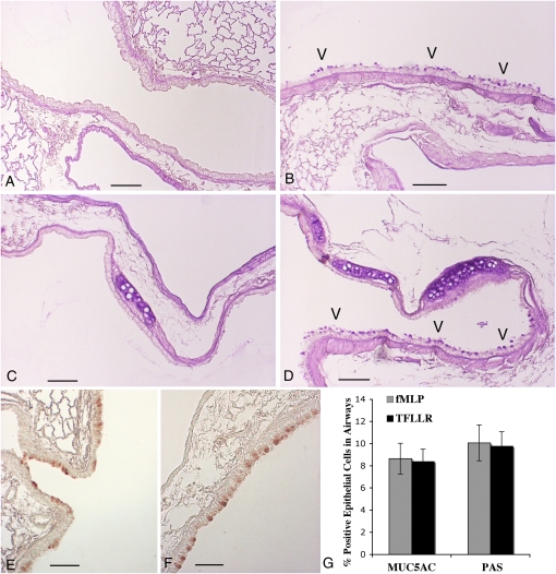
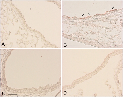
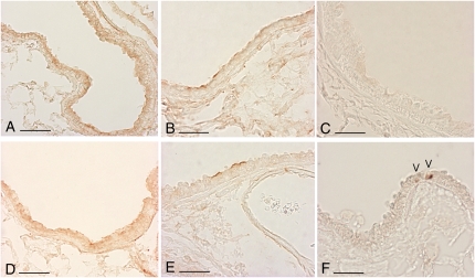
The morphological features of chronic obstructive pulmonary disease in man include emphysema and chronic bronchitis associated with mucus hypersecretion. These alterations can be induced in mice by a single intratracheal instillation of N-formyl-L-methionyl-L-leucyl-L-phenylalanine (fMLP), a chemoattractant and degranulating agent for neutrophils. The mechanisms underlying excessive mucus production and, in particular, goblet cell hyperplasia/metaplasia in chronic obstructive pulmonary disease remain poorly understood. The proteinase-activated receptors (PARs) are widely recognized for their modulatory properties during inflammation. In this study, we examined whether PAR-1 contributes to inflammation and lung damage induced by fMLP by comparing the response of PAR-1-deficient (PAR-1(-/-)) mice with that of wild-type (WT) mice. Mice were killed at various time points after fMLP instillation (200 microg/50 microl). WT mice developed emphysema and goblet cell metaplasia. The onset of pulmonary lesions was preceded by an increase in thrombin immunoreactivity in bronchial airways and alveolar tissue. This was followed by a decrease in PAR-1 immunoreactivity, and by an increase in IL-13 immunostaining on the luminal surface of airway epithelial cells. In PAR-1(-/-) mice, fMLP administration induced similar responses in terms of inflammation and emphysema, but these mice were protected from the development of goblet cell metaplasia. The involvement of PAR-1 in airway epithelial cell transdifferentiation was confirmed by demonstrating that intratracheal instillation of the selective PAR-1 agonist (TFLLR) induced goblet cell metaplasia in the airways of WT mice only. These data suggest that emphysema and goblet cell metaplasia occur independently, and that PAR-1 signaling through IL-13 stimulation may play an important role in inducing goblet cell metaplasia.


Figures

Figure 1.
Figure 1.
N-formyl-L-methionyl-L-leucyl-L-phenylalanine (fMLP) instillation induces neutrophil infiltration and airspace enlargement in wild-type (WT) and PAR-1–deficient (PAR-1−/−) mice. Histological sections from lungs of C57BL/6 and PAR-1−/− mice at 24 hours and 21 days after fMLP instillation are compared with their respective saline-instilled control mice. Representative hematoxylin and eosin (H&E) lung sections of C57BL/6 (A) and PAR-1−/− mice (D) show consistent neutrophil infiltration into alveolar spaces 24 hours after fMLP instillation. Representative sections of lungs from C57 BL/6 (B) and PAR-1−/− (E) mice 21 days after fMLP instillation are characterized by diffuse areas of air space enlargement with destruction of alveolar septa. No differences in lung histological appearance are found between saline-instilled WT and PAR-1−/− mice (C and F). H&E stain; scale bar, 170 μm.
Figure 2.
Figure 2.
PAR-1−/− mice are protected from fMLP-induced goblet cell metaplasia. Goblet cell metaplasia (arrowheads) is appreciable in epithelium of large- (A) and middle-sized bronchi (B) of WT C57BL/6 mice 21 days after fMLP instillation. Areas of goblet cell metaplasia are found in some C57BL/6 mice as early as 3 days after fMLP treatment (C). Bronchial sections of airway epithelium from PAR-1−/− mice at 21 days after fMLP instillation are reported in D and E. As in saline control mice (F and G), no periodic acid-Schiff (PAS)–positive cells are present in these sections. PAS stain; scale bar, 150 μm.
Figure 3.
Figure 3.
PAR-1 immunoreactivity decreases after fMLP instillation. Immunohistochemical localization of PAR-1 is shown on alveolar and bronchiolar epithelial cells (A), on the epithelium of middle- (B) and large-size (C) bronchi, and on endothelial cells within blood vessels (B and C) from a control C57BL/6 mouse. In these sections, PAR-1 is consistently expressed throughout the lung parenchyma and airways. In contrast, PAR-1 immunoreactivity associated with the parenchyma (D) and airway epithelium (E and F) is dramatically decreased on Day 1 after fMLP instillation. An almost complete recovery in PAR-1 immunoreactivity is observed throughout the pulmonary structures at 7 days after fMLP instillation (G and H). Scale bar, 100 μm.
Figure 4.
Figure 4.
Thrombin immunoreactivity is increased after fMLP exposure. Immunohistochemical localization of thrombin in lung sections from control (A and B) and fMLP-instilled (C and D) C57BL/6 mice. Only minimal immunoreactivity is observed within the capillary vessels (A) and around the bronchial structures (B). At 1 day after instillation of fMLP, there is increased staining for thrombin in alveolar exudates (C) and on bronchial epithelial cell surfaces (D). Scale bar, 75 μm. (E) Representative Western blot analysis of thrombin in lung homogenates from C57BL/6 mice at 0, 1, and 3 days after fMLP instillation. Active thrombin is appreciable in lung homogenates at 1 and 3 days after fMLP instillation.
Figure 5.
Figure 5.
PAR-1 activation induces goblet cell metaplasia in WT mice. PAS staining (AD) of sections demonstrates that goblet cell metaplasia (arrowheads) is appreciable within the epithelium of middle- (B) and large-sized (D) bronchi of WT C57BL/6 mice 14 days after fMLP instillation. Middle-sized and large bronchial sections of airway epithelium from a saline control WT mouse at 14 days after treatment are reported in A and C. No PAS-positive cells are present in these sections. MUC5AC immunostaining (E and F) of sections confirms goblet cell metaplasia in fMLP- (E) and TFLLR-instilled (F) mice. Scale bar, 150 μm. (G) Quantitation of MUC5AC- and PAS-positive cells in airways from fMLP- and TFLLR-instilled mice at 14 days after treatment. Data are expressed as a percentage of PAS- or MUC5AC-positive airway epithelial cells. Each bar represents the mean ± SD (n = 8 intrapulmonary airway sections).
Figure 6.
Figure 6.
IL-13 immunoreactivity increases after fMLP exposure. IL-13 immunoreactivity was consistently associated with the airway epithelium of WT mice after fMLP treatment from Day 1 onwards. IL-13 immunoreactivity is associated with the luminal surface of epithelial cells within the bronchi (B) of WT mice on Day 1 after fMLP instillation (arrowheads). IL-13 immunoreactivity is absent in airway epithelium of PAR-1−/− mice at Day 1 after fMLP instillation (D). (A and C) The airway epithelium for saline control WT and PAR-1−/− mice. IL-13 was undetectable in these sections. Scale bar, 150 μm.
Figure 7.
Figure 7.
Epidermal growth factor receptor (EGFR) and phosphorylated EGFR (pEGFR) immunoreactivity is unchanged by fMLP exposure. (A, B, D, and E) Representative sections immunostained for EGFR from an untreated WT mouse (A), a WT mouse at Day 1 after fMLP instillation (B), an untreated PAR-1−/− mouse (D), and a PAR-1−/− mouse at Day 1 after fMLP instillation. EGFR staining was similar for all groups of mice at all time points examined. Scale bar, 150 μm. (C) A representative section immunostained for activated EGFR (pEGFR) for WT mice on Day 7 after fMLP treatment. pEGFR immunostaining was similar for all groups of mice at all time points examined. However, positive pEGFR nuclear staining was occasionally found in a very small number of cells in mice belonging to the various experimental groups on Day 7 after fMLP challenge. (F) pEGFR nuclear staining (arrowheads) in a representative section for PAR-1−/− mice on Day 7 after fMLP treatment. Scale bar, 150 μm.

Similar articles

Cited by

References

    1. Saetta M, Turato G, Lupi F, Fabbri LM. Inflammation in the pathogenesis of chronic obstructive pulmonary disease. In: Voelkel NF and MacNee W, editors. Chronic obstructive lung disease. Hamilton, ON, Canada: BC Decker; 2002. pp. 114–126.
    1. Minematsu N, Shapiro SD. To live and die in the la (lung airway): mode of neutrophil death and progression of chronic obstructive pulmonary disease. Am J Respir Cell Mol Biol 2007;37:129–130. - PubMed
    1. Tetley TD. Inflammatory cells and chronic obstructive pulmonary disease. Curr Drug Targets Inflamm Allergy 2005;4:607–618. - PubMed
    1. Nadel JA. Mucus and mucus-secreting cells. In: Voelkel NF and MacNee W, editors. Chronic obstructive lung disease. Hamilton, ON, Canada: BC Decker; 2002. pp.161–174.
    1. Barnes PJ. Mediators of chronic obstructive pulmonary disease. Pharmacol Rev 2004;56:515–548. - PubMed

Publication types

MeSH terms