. 2009 Apr;119(4):899-910.
doi: 10.1172/JCI37771. Epub 2009 Mar 23.

Loss of serum response factor in keratinocytes results in hyperproliferative skin disease in mice

Affiliations

Loss of serum response factor in keratinocytes results in hyperproliferative skin disease in mice

Heidi Koegel et al. J Clin Invest. 2009 Apr.

Abstract

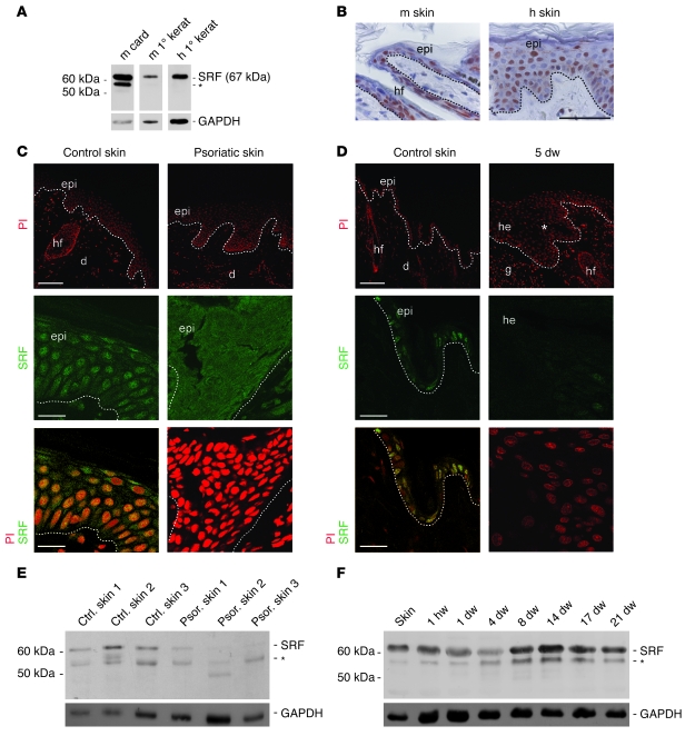
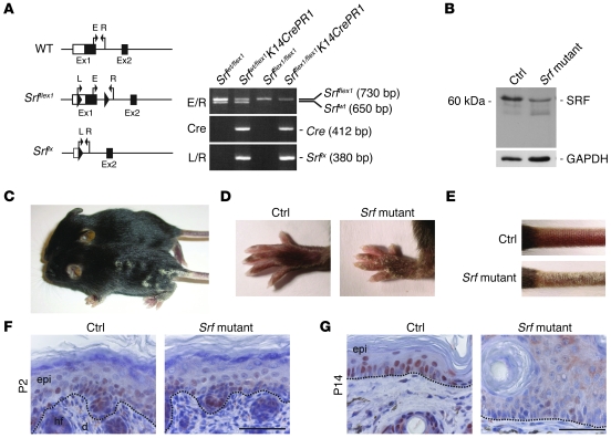
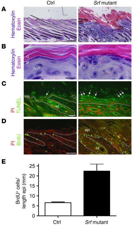
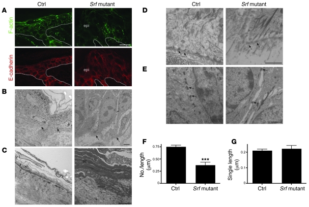
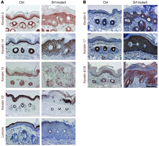
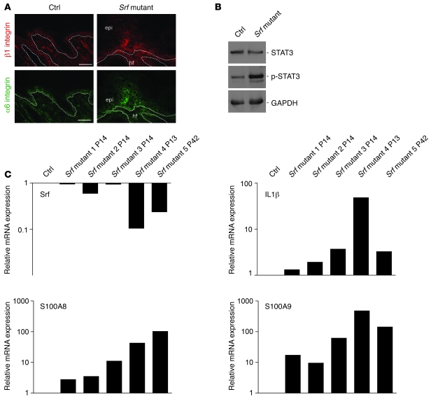
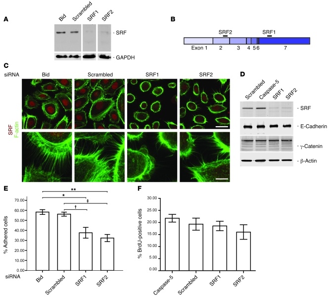
The transcription factor serum response factor (SRF) plays a crucial role in the development of several organs. However, its role in the skin has not been explored. Here, we show that keratinocytes in normal human and mouse skin expressed high levels of SRF but that SRF expression was strongly downregulated in the hyperproliferative epidermis of wounded and psoriatic skin. Keratinocyte-specific deletion within the mouse SRF locus during embryonic development caused edema and skin blistering, and all animals died in utero. Postnatal loss of mouse SRF in keratinocytes resulted in the development of psoriasis-like skin lesions. These lesions were characterized by inflammation, hyperproliferation, and abnormal differentiation of keratinocytes as well as by disruption of the actin cytoskeleton. Ultrastructural analysis revealed markedly reduced cell-cell and cell-matrix contacts and loss of cell compaction in all epidermal layers. siRNA-mediated knockdown of SRF in primary human keratinocytes revealed that the cytoskeletal abnormalities and adhesion defects were a direct consequence of the loss of SRF. In contrast, the hyperproliferation observed in vivo was an indirect effect that was most likely a consequence of the inflammation. These results reveal that loss of SRF disrupts epidermal homeostasis and strongly suggest its involvement in the pathogenesis of hyperproliferative skin diseases, including psoriasis.


Figures

Figure 1
Figure 1. Downregulation of SRF expression in hyperproliferative epidermis.
(A) SRF expression in murine (m) and human (h) primary (1°) keratinocytes (kerat) and in murine cardiac muscle (card) shown by Western blotting using a rat anti-SRF mAb (2C5). (B) Immunohistochemical analysis of sections from mouse and human skin using the 2C5 antibody shows nuclear localization of SRF in the epidermis (epi). hf, hair follicle. Scale bar: 50 μm. (C) Immunofluorescence stainings of SRF (green, middle and lower panels) show nuclear localization of SRF in epidermal keratinocytes of a healthy patient. Nuclear SRF was not detected in lesional skin of a psoriatic patient. PI-stained sections are shown at low magnification in the upper panels. Representative biopsies (from 5 healthy and 5 psoriatic patients) are shown. Control skin, yellow from double staining with propidium iodide (PI), red. d, dermis. Scale bars: 100 μm (upper panels); 20 μm (middle and lower panels). (D) Immunofluorescence stainings of SRF (middle panels and, in combination with PI, lower panels) show nuclear localization in keratinocytes of normal mouse skin but reduced staining in the hyperproliferative epithelium of 5-day wounds (5 dw). PI-stained sections are shown at low magnification in the upper panels. Asterisk indicates area of hyperproliferative epithelium at the wound edge shown at higher magnification in the lower panels. g, granulation tissue; he, hyperproliferative epithelium. Scale bars: 100 μm (upper panels); 20 μm (middle and lower panels). Dotted lines indicate basement membrane. (E and F) Western blot analysis of SRF using lysates from 3 healthy patients (ctrl) and affected skin of 3 psoriatic (psor) patients (E) or lysates of normal mouse skin and excisional wounds (F). (A, E, and F) Asterisks denote an additional band that is recognized by the anti-SRF antibody. GAPDH was used as a loading control.
Figure 2
Figure 2. Spontaneous recombination of the floxed Srf allele in Srfflex1/flex1K14CrePR1 mice.
(A) Schematic illustration of Srf alleles and genotyping of Srfflex1/flex1 × SrfWT/flex1K14CrePR1 offspring: Srfflex1/flex1K14CrePR1 (Srf mutant) mice were identified by a 730-bp fragment (Srfflex1), a 412-bp fragment (Cre recombinase), and the absence of the 650-bp fragment that identifies the WT allele (SrfWT). A 380-bp fragment was amplified from the recombined Srf allele (Srflx). E/R and L/R indicate primer pairs described in ref. . (B) A representative Western blot of total back skin lysates showing reduced SRF protein levels in the skin of Srfflex1/flex1K14CrePR1 mice at P14 compared with skin of control littermates. GAPDH was used as a loading control. (CE) Macroscopic appearance of control and Srf mutant mice at P14. Note the scaly lesions on the back (C), the paws (D), and the tail (E). (F) Immunohistochemical analysis of SRF in back skin from Srf mutant and control mice at P2 shows comparable SRF expression patterns. (G) At P14, nuclear SRF staining is markedly reduced in the epidermis of Srfflex1/flex1K14CrePR1 mice. (F and G) Dotted lines indicate the basement membrane. Scale bars: 50 μm. Sections were counterstained with hematoxylin.
Figure 3
Figure 3. Loss of SRF expression in keratinocytes leads to hyperproliferative epidermis.
(A and B) H&E staining of back skin sections of control and Srf mutant mice shows epidermal hyperthickening and parakeratosis in skin lesions of Srf mutant mice. At high magnification, the presence of a granular layer was still seen in most of the mutant mice (B). Scale bar: 10 μm. cl, cornified layer; gl, granular layer; nu, nucleus. (C) TUNEL staining in combination with PI shows an increased number of apoptotic keratinocytes in lesional skin of Srf mutant mice (indicated by arrows). (D) Immunofluorescence analysis in combination with PI shows an increased number of proliferating keratinocytes by BrdU staining. Dotted lines indicate the basement membrane. (E) SRF mutant epidermis shows an increased number of BrdU-positive cells compared with control epidermis. n = 3 per genotype. Error bars indicate mean ± SEM. Scale bars: 200 μm (A); 10 μm (B); 20 μm (C); 100 μm (D).
Figure 4
Figure 4. Disruption of the actin cytoskeleton and reduced cell-cell and cell-matrix contacts in Srf mutant mice.
(A) Phalloidin-FITC staining of skin sections of control and Srf mutant mice at P14 shows markedly reduced levels of polymerized actin (F-actin) in lesions of Srf mutant skin (upper panels). Immunofluorescence staining for E-cadherin shows membrane localization in skin of control and Srf mutant mice (lower panels). Dotted lines indicate the basement membrane. Scale bar: 20 μm. (BE) Ultrastructural analysis of control and Srf mutant epidermis at P14. (B) Keratinocytes in the basal cell layer of lesional Srf mutant epidermis are strongly enlarged and lack most cell-cell contacts compared with control skin, in which basal keratinocytes are small and densely packed. Arrows indicate the basement membrane. (C) Stratum corneum (sc) of lesional Srf mutant epidermis is thicker and less compact than stratum corneum of control epidermis. (D) Epidermal lesions of Srf mutant skin show areas with defective hemidesmosomes (arrows). Note that collagen bundles below the basement membrane are absent in Srf mutant skin. (E) Desmosomes (arrows) are markedly reduced in Srf mutant skin, and cell-cell contacts are largely absent. (F and G) Morphometric analysis confirmed a distinct reduction of the number of desmosomes (F), while no change of the mean length of single desmosomes could be found (G). Error bars indicate SD. Scale bars: 5 μm (B and C); 1 μm (D and E). ***P ≤ 0.001.
Figure 5
Figure 5. Keratinocyte differentiation is impaired in Srf mutant epidermis.
(A and B) Immunohistochemical analysis of tail-skin sections from control and Srf mutant mice (P11–P15) for the expression of epidermal differentiation markers. (A) Expression of K14, K15, and K5 was reduced and their localization was irregular in Srf mutant tail skin. Expression of the differentiation markers K10 and loricrin was delayed and reduced compared with that in control skin. (B) Srf mutant mice show abnormal interfollicular expression of K6, K17, and K8/K18. Sections were counterstained with hematoxylin. Scale bar: 100 μm.
Figure 6
Figure 6. Skin lesions of Srf mutant mice reveal markers of inflammatory skin disease.
(A) Immunofluorescence analysis of skin sections shows high suprabasal expression of the β1 integrin (upper panels) and α-integrin (lower panels) subunits in Srf mutant skin. Scale bars: 20 μm. Dotted lines indicate the basement membrane. (B) p-STAT3 levels are markedly increased in total skin lysates from Srf mutant mice. GAPDH was used as a loading control. (C) Relative mRNA levels of Srf, IL-1β, S100A8, and S100A9 in 5 individual Srf mutant mice are shown (1–3, P14; 4, P13: severe scaling phenotype; 5, P42: slowly progressing phenotype). Reduced Srf expression correlates with increased IL-1β, S100A8 and S100A9 mRNA levels. RNA levels of a control littermate are set as 1. mRNA of the housekeeping gene Gapdh was used for normalization. PCR analysis was performed in duplicate, and the average of both values is shown. Each result was reproduced in an independent experiment.
Figure 7
Figure 7. siRNA-mediated downregulation of SRF in primary human keratinocytes disrupts the actin cytoskeleton and affects cell adhesion but not proliferation.
(A) SRF protein is efficiently downregulated in primary human keratinocytes as shown by Western blot analysis. Scrambled siRNA and siRNA against Bid served as controls. GAPDH was used as a loading control. (B) Schematic representation of SRF mRNA. Black bars indicate siRNA-binding sites. (C) Immunofluorescence analysis of SRF (red) shows reduced nuclear staining in SRF siRNA–treated keratinocytes. Costaining with phalloidin-FITC (green) revealed reduced cell size (see upper panels) and less filopodia (see lower panels) in SRF1 and SRF2 siRNA–treated keratinocytes. Scale bars: 20 μm (upper panels); 5 μm (lower panels). (D) Western blot analysis showing expression of SRF, E-cadherin, γ-catenin (plakoglobin), and β-actin in keratinocytes treated with scrambled or SRF siRNAs. siRNA against an unrelated protein (caspase-5) was used as an additional control. (E) Adhesion efficiency of SRF siRNA–treated keratinocytes is significantly reduced (n = 3). Error bars show mean ± SEM, *P = 0.021 (Bid/SRF1); P = 0.038 (scrambled/SRF1); **P = 0.005 (Bid/SRF2); P = 0.009 (scrambled/SRF2), ANOVA with Bonferroni’s post hoc test. (F) BrdU incorporation was analyzed in keratinocytes that had been treated for 3 days with SRF siRNAs or with control siRNAs (n = 3 wells per treatment group; 10 microscopic fields per dish were counted). Error bars show mean ± SEM. No significant difference was detected using ANOVA with Bonferroni post hoc test.

Similar articles

Cited by

References

    1. Fuchs E., Raghavan S. Getting under the skin of epidermal morphogenesis. Nat. Rev. Genet. 2002;3:199–209. doi: 10.1038/nrg758. - DOI - PubMed
    1. Shore P., Sharrocks A.D. The MADS-box family of transcription factors. Eur. J. Biochem. 1995;229:1–13. doi: 10.1111/j.1432-1033.1995.tb20430.x. - DOI - PubMed
    1. Norman C., Runswick M., Pollock R., Treisman R. Isolation and properties of cDNA clones encoding SRF, a transcription factor that binds to the c-fos serum response element. Cell. 1988;55:989–1003. doi: 10.1016/0092-8674(88)90244-9. - DOI - PubMed
    1. Treisman R. Identification of a protein-binding site that mediates transcriptional response of the c-fos gene to serum factors. Cell. 1986;46:567–574. doi: 10.1016/0092-8674(86)90882-2. - DOI - PubMed
    1. Sun Q., et al. Defining the mammalian CArGome. Genome Res. 2006;16:197–207. doi: 10.1101/gr.4108706. - DOI - PMC - PubMed

Publication types

MeSH terms