

Prevention of hypoxia by myoglobin expression in human tumor cells promotes differentiation and inhibits metastasis
- PMID: 19307731
- PMCID: PMC2662552
- DOI: 10.1172/JCI36579
Prevention of hypoxia by myoglobin expression in human tumor cells promotes differentiation and inhibits metastasis
Abstract
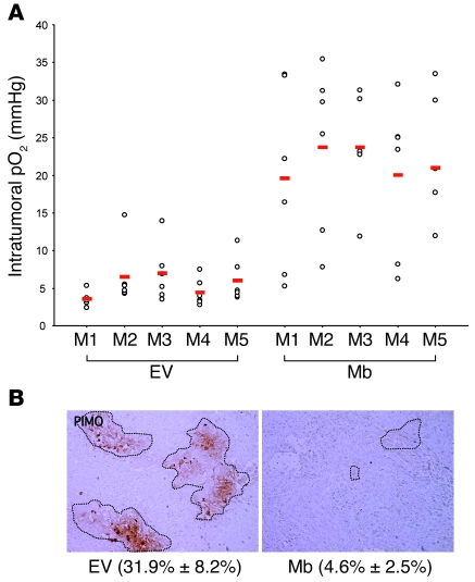
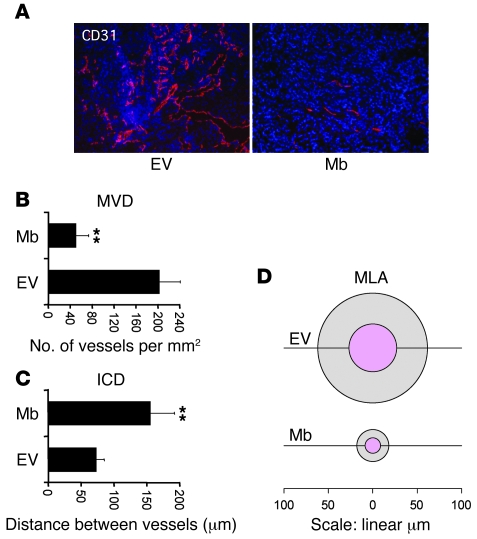
As a tumor grows, it requires increased amounts of oxygen. However, the tumor blood vessels that form to meet this demand are functionally impaired, leading to regions of hypoxia within the tumor. Such hypoxia is one of the hallmarks of malignancy and is thought to promote a number of tumorigenic properties. Here, we sought to determine how tumors without hypoxia would progress by engineering A549 human lung carcinoma cells to ectopically express myoglobin (Mb), a multifunctional heme protein that specializes in oxygen transport, storage, and buffering. Mb expression prevented the hypoxic response in vitro and delayed tumor engraftment and reduced tumor growth following xenotransplantation into mice. Experimental tumors expressing Mb displayed reduced or no hypoxia, minimal HIF-1alpha levels, and a homogeneously low vessel density. Mb-mediated tumor oxygenation promoted differentiation of cancer cells and suppressed both local and distal metastatic spreading. These effects were primarily due to reduced tumor hypoxia, because they were not observed using point-mutated forms of myoglobin unable to bind oxygen and they were abrogated by expression of a constitutively active form of HIF-1alpha. Although limited to xenograft models, these data provide experimental proof of the concept that hypoxia is not just a side effect of deregulated growth but a key factor on which the tumor relies in order to promote its own expansion.
Figures


Comment in
-
Myoglobin tames tumor growth and spread.J Clin Invest. 2009 Apr;119(4):766-8. doi: 10.1172/jci38796. J Clin Invest. 2009. PMID: 19348046 Free PMC article.
Similar articles
-
Myoglobin tames tumor growth and spread.J Clin Invest. 2009 Apr;119(4):766-8. doi: 10.1172/jci38796. J Clin Invest. 2009. PMID: 19348046 Free PMC article.
-
Hypoxia induces tumor aggressiveness and the expansion of CD133-positive cells in a hypoxia-inducible factor-1α-dependent manner in pancreatic cancer cells.Pathobiology. 2011;78(4):181-92. doi: 10.1159/000325538. Epub 2011 Jul 19. Pathobiology. 2011. PMID: 21778785
-
NRF2 blockade suppresses colon tumor angiogenesis by inhibiting hypoxia-induced activation of HIF-1α.Cancer Res. 2011 Mar 15;71(6):2260-75. doi: 10.1158/0008-5472.CAN-10-3007. Epub 2011 Jan 28. Cancer Res. 2011. PMID: 21278237
-
The role of the hypoxia-inducible factor in tumor metabolism growth and invasion.Bull Cancer. 2006 Aug;93(8):E73-80. Bull Cancer. 2006. PMID: 16935775 Review.
-
Hypoxia and Metabolism in Metastasis.Adv Exp Med Biol. 2019;1136:87-95. doi: 10.1007/978-3-030-12734-3_6. Adv Exp Med Biol. 2019. PMID: 31201718 Review.
Cited by
-
Can we negotiate with a tumor?PLoS One. 2014 Aug 1;9(8):e103834. doi: 10.1371/journal.pone.0103834. eCollection 2014. PLoS One. 2014. PMID: 25084359 Free PMC article.
-
The Distinct Gene Regulatory Network of Myoglobin in Prostate and Breast Cancer.PLoS One. 2015 Nov 11;10(11):e0142662. doi: 10.1371/journal.pone.0142662. eCollection 2015. PLoS One. 2015. PMID: 26559958 Free PMC article.
-
Treatment Strategies Considering Micro-Environment and Clonal Evolution in Multiple Myeloma.Cancers (Basel). 2021 Jan 8;13(2):215. doi: 10.3390/cancers13020215. Cancers (Basel). 2021. PMID: 33435539 Free PMC article. Review.
-
Metabolic regulation of oxygen and redox homeostasis by p53: lessons from evolutionary biology?Free Radic Biol Med. 2012 Sep 15;53(6):1279-85. doi: 10.1016/j.freeradbiomed.2012.07.026. Epub 2012 Jul 25. Free Radic Biol Med. 2012. PMID: 22841759 Free PMC article. Review.
-
Pro-Apoptotic and Anti-Invasive Properties Underscore the Tumor-Suppressing Impact of Myoglobin on a Subset of Human Breast Cancer Cells.Int J Mol Sci. 2022 Sep 29;23(19):11483. doi: 10.3390/ijms231911483. Int J Mol Sci. 2022. PMID: 36232784 Free PMC article.
References
Publication types
MeSH terms
Substances
LinkOut - more resources
Full Text Sources
Other Literature Sources
Research Materials
Miscellaneous
