. 2009 May 31;41(5):341-8.
doi: 10.3858/emm.2009.41.5.038.

ICAM-1/LFA-1 interaction contributes to the induction of endothelial cell-cell separation: implication for enhanced leukocyte diapedesis

Affiliations

ICAM-1/LFA-1 interaction contributes to the induction of endothelial cell-cell separation: implication for enhanced leukocyte diapedesis

Hyun Wee et al. Exp Mol Med. .

Abstract

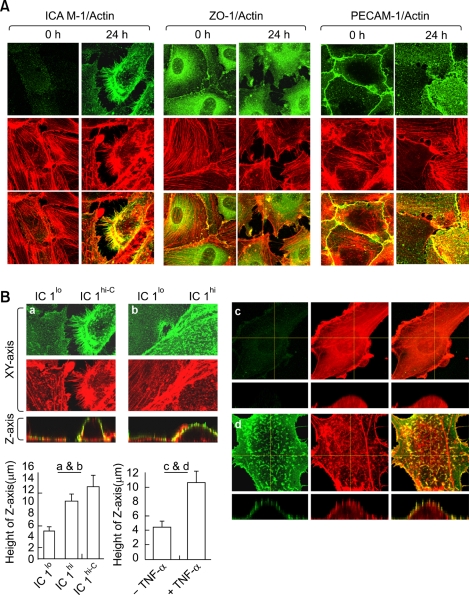
The basic route and mechanism for diapedesis has not yet to be fully defined. Here we present evidence that "cell-cell separation" between endothelial cells (ECs) may provide a route for leukocyte diapedesis. We unexpectedly found that extensive interaction between peripheral blood leukocytes and ECs that were activated by TNF-alpha induced the opening of EC contacts and, surprisingly, resulted in cell-cell separation. This event was specific to the intercellular adhesion molecules-1 (ICAM-1)/leukocyte function- associated antigen-1 interaction, as demonstrated by the following: (1) ICAM-1 expression correlated with increased EC contraction; and (2) the blocking of ICAM-1 selectively inhibited EC separation. Thus, we suggest that "cell-cell separation" could be a mechanism for diapedesis in situations that may require massive leukocyte infiltration.


Figures

Figure 1
Figure 1
Dynamic behavior of HUVECs during the interaction with leukocytes under shear-flow condition. (A) Experimental scheme for live-cell dynamic imaging as described in Methods. (B) Dynamic behavior of endothelial cells under a shear-flow condition. Upper panels show time-lapse images of non-activated HUVECs with and without interaction with PBLs (2 × 10⁵ cells/200 µl). Lower panels show time-lapse images of TNF-α activated HUVECs under the same experimental conditions. (C) Increased number of PBLs (1 × 10⁶ cells/200 µl) were flowed on TNF-α activated HUVECs and resulted in "cell-cell separation" between HUVECs.
Figure 2
Figure 2
Effects of ICAM-1 expression on EC height and contractility. (A) The monolayers of HUVECs were incubated with TNF-α for 0 h or 24 h at 37℃. Cells were fixed and stained with ICAM-1 (R6.5; green), ZO-1 (green), PECAM-1 (green) and actin (phallodin-TRITC; red) and subjected to confocal microscopy with reconstitution in the z-axis. (B) The expression level of the ICAM-1 varied in TNF-α stimulated HUVECs for 24 h. The EC height of EC expressing low level of ICAM-1 (IC1lo), high level of ICAM-1 (IC1hi) and contracted EC expressing high level of (IC1hi-C) was analyzed, respectively (a and b). The EC height of HUVECs stimulated or unstimulated with TNF-α for 24 h was measured, respectively (c and d). The crossline (yellow) represents the site for the indicated orthogonal view. EC height was calculated by the sum of the z-axes in an orthogonal view (height of combined Z-sections) using FLUOVIEW software.
Figure 3
Figure 3
ICAM-1/LFA-1 interaction is essential for leukocyte-induced cell-cell separation between ECs. PBLs (5 × 10⁵ cells) stimulated with SDF-α were incubated on monolayers of HUVECs with or without TNF-α stimulation and monitored by time-lapse microscopy. The cells were treated with R6.5 antibody (10 µg/ml) to block the interaction of ICAM-1 with LFA-1. Selected images are shown at 0, 10, 30, 50 and 60 min of incubation from a representative experiment. White dot lines represent the border line of cell membranes and the arrow heads indicate a change of direction between contraction and respreading.

References

    1. Cinamon G, Shinder V, Alon R. Shear forces promote lymphocyte migration across vascular endothelium bearing apical chemokines. Nat Immunol. 2001;2:515–522. - PubMed
    1. Clark PR, Manes TD, Pober JS, Kluger MS. Increased ICAM-1 expression causes endothelial cell leakiness, cytoskeletal reorganization and junctional alterations. J Invest Dermatol. 2007;127:762–774. - PubMed
    1. Feng D, Nagy JA, Pyne K, Dvorak HF, Dvorak AM. Neutrophils emigrate from venules by a transendothelial cell pathway in response to FMLP. J Exp Med. 1998;187:903–915. - PMC - PubMed
    1. Gahmberg CG, Tolvanen M, Kotovuori P. Leukocyte adhesion--structure and function of human leukocyte beta2-integrins and their cellular ligands. Eur J Biochem. 1997;245:215–232. - PubMed
    1. Godaly G, Bergsten G, Hang L, Fischer H, Frendeus B, Lundstedt AC, Samuelsson M, Samuelsson P, Svanborg C. Neutrophil recruitment, chemokine receptors, and resistance to mucosal infection. J Leukoc Biol. 2001;69:899–906. - PubMed

Publication types

MeSH terms

Substances

LinkOut - more resources