. 2009;4(3):e4994.
doi: 10.1371/journal.pone.0004994. Epub 2009 Mar 24.

Discovery and characterization of novel vascular and hematopoietic genes downstream of etsrp in zebrafish

Affiliations

Discovery and characterization of novel vascular and hematopoietic genes downstream of etsrp in zebrafish

Gustavo A Gomez et al. PLoS One. 2009.

Abstract

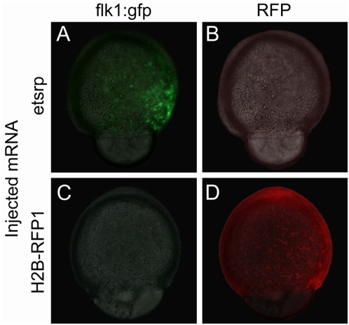
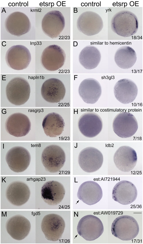
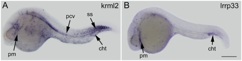
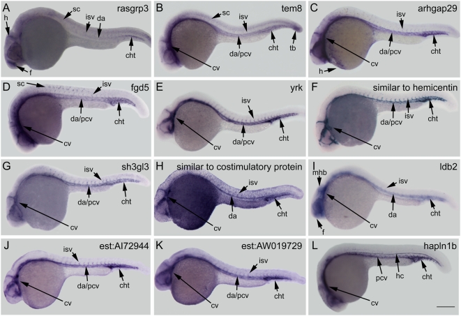
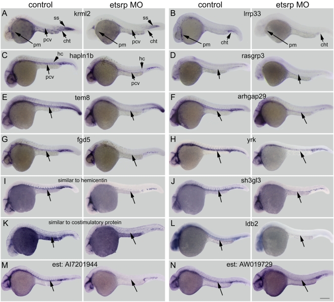
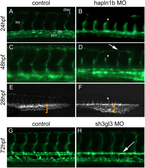
The transcription factor Etsrp is required for vasculogenesis and primitive myelopoiesis in zebrafish. When ectopically expressed, etsrp is sufficient to induce the expression of many vascular and myeloid genes in zebrafish. The mammalian homolog of etsrp, ER71/Etv2, is also essential for vascular and hematopoietic development. To identify genes downstream of etsrp, gain-of-function experiments were performed for etsrp in zebrafish embryos followed by transcription profile analysis by microarray. Subsequent in vivo expression studies resulted in the identification of fourteen genes with blood and/or vascular expression, six of these being completely novel. Regulation of these genes by etsrp was confirmed by ectopic induction in etsrp overexpressing embryos and decreased expression in etsrp deficient embryos. Additional functional analysis of two newly discovered genes, hapln1b and sh3gl3, demonstrates their importance in embryonic vascular development. The results described here identify a group of genes downstream of etsrp likely to be critical for vascular and/or myeloid development.


Conflict of interest statement

Competing Interests: The authors have declared that no competing interests exist.

Figures

Figure 1
Figure 1. Early ectopic expression of transgenic flk1:gfp is induced by etsrp over-expression.
(A) Injection of 75 pg of etsrp mRNA at one-cell stage results in the ectopic induction of flk1:gfp before the end of gastrulation. (B) No red fluorescence is observed in etsrp mRNA injected flk1:gfp transgenic embryos. (C) Injection of 500 pg of control H2B-RFP1 mRNA does not result in the early induction of flk1:gfp similar to uninjected embryos (not shown). (D) Uniform H2B-RFP1 expression in a H2B-RFP1 mRNA injected embryo. Panels A and C are composite images of light transmitted images merged with the green fluorescent channel, while panels B and D are composites of light transmitted images merged with the red fluorescent channel.
Figure 2
Figure 2. Ectopic induction of microarray identified genes by etsrp overexpression.
Ectopic gene expression was examined at 80% epiboly to tailbud stages in flk1:gfp transgenic embryos injected with 30 pg etsrp-mcherry DNA at the one cell stage. Embryos exhibiting both red and green fluorescence were selected for analysis. Representative control embryos are on the left and etsrp overexpressing (OE) embryos are on the right side of each panel. Represented genes: (A) krml2; (B) yrk; (C) lrrp33; (D) similar to hemicentin; (E) hapln1b; (F) sh3gl3; (G) rasgrp3; (H) similar to costimulatory protein; (I) tem8; (J) ldb2; (K) arhgap23; (L) est:AI721944; (M) fgd5; and (N) est:AW019729. Note that there is a low level of endogenous expression in the control embryos for the two EST's, L and N, at the polster (arrows). Ratios in bottom right hand corner in panels represent the number of embryos with ectopic induction of total embryos processed and scored in the injected groups; control embryos never displayed ectopic induction. All embryos are in lateral view, and those at tail bud stage are oriented with anterior to the left. Scale bar: 250 µm.
Figure 3
Figure 3. Expression of genes identified in primitive myeloid blood cells.
Gene expression was examined at 24–28 hours post fertilization by whole mount in situ hybridization in wildtype embryos. (A) krml2 is expressed in primitive myeloid cells (dispersed cells labeled throughout yolk and head) in the posterior cardinal vein, caudal hematopoietic tail region, and somites. (B) Lrrp33 is expressed in primitive myeloid cells and in the caudal hematopoietic tissue Abbreviations: pm, primitive myeloid; pcv, posterior cardinal vein; cht, caudal hematopoietic tail region; ss, somites. Scale bar: 250 µm.
Figure 4
Figure 4. Expression of genes identified in vascular endothelial cells.
Gene expression was examined at 24–28 hours post fertilization by whole mount in situ hybridization in wildtype embryos. (A) rasgrp3 is expressed weakly in the forebrain, hindbrain, spinal cord, intersomitic vessels, dorsal aorta, and caudal hematopoietic tail region. (B) tem8 is expressed highly in the cranial vasculature, the dorsal aorta and posterior cardinal vein, tailbud and weakly in the intersomitic vessels and spinal cord. (C) arhgap29 is expressed in the cranial vessels, dorsal aorta, posterior cardinal vein, intersomitic vessels, caudal hematopoetic tail region, and hatching gland. (D) fgd5 is expressed in the cranial vasculature, dorsal aorta, posterior cardinal vein, intersomitic vessels, and caudal hematopoetic tail region. (E, F, and G) yrk, similar to hemicentin, and sh3gl3 are all expressed in the cranial vasculature, dorsal aorta, posterior cardinal vein, intersegmental vessels, and caudal vascular hematopoietic tail region. (H) similar to costimulatory protein is expressed in the cranial vasculature, dorsal aorta, intersomitic vessels, and caudal hematopoietic tail region; additional expression is present in the ectoderm layer throughout the embryo. (I) ldb2 is expressed in the forebrain, midbrain-hindbrain boundary, dorsal aorta, intersomitic vessels, and caudal hematopoietic tail region. (J and K) Both est:AI721944 and est:AW019729 are expressed in the cranial vasculature, dorsal aorta, posterior cardinal vein, intersomitic vessels, and caudal hematopoietic tail region. (L) hapln1b is expressed in the cranial vasculature, posterior cardinal vein, caudal hematopoietic tail region, and hypochord. Abbreviations: f, forebrain; h, hindbrain; sc, spinal cord; cv, cranial vasculature; da, dorsal aorta; pcv, posterior cardinal vein; isv, intersomitic vessels; cht, caudal hematopoietic tail region; tb, tail bud; h, hatching gland; and hc, hypochord. Scale bar: 250 µm.
Figure 5
Figure 5. Morpholino knockdown of etsrp reduces expression of genes identified by etsrp overexpression microarray analysis.
Embryos were injected with a combination of two etsrp targeting morpholinos (4 ng each) and gene expression was examined by in situ hybridization at 24–26 hours post fertilization. Representative control embryos are on the left of each panel and etsrp morpholino (MO) injected embryos are on the right. (A) krml2 is reduced in primitive myeloid cells, posterior cardinal vein, and caudal hematopoietic tail region by etsrp knockdown. (B) lrrp33 expression is absent in myeloid cells and reduced in the caudal hematopoietic tail region. (C) hapln1b expression is lost in the posterior cardinal vein when etsrp is knocked down. (D–N) Etsrp knockdown causes a significant decrease of gene expression, most pronounced in the trunk (unlabeled arrows). (D) rasgfp3; (E) tem8; (F) arhgap29; (G) fgd5; (H) yrk; (I) similar to hemicentin; (J) sh3gl3; (K) similar to costimulatory protein; (L) ldb2; (M) est:AI7201944; and (N) est:AW019729. Note that non-hematovascular tissues such as somites in (A) and hypochord in (C) are not affected by etsrp knockdown. Abbreviations: pm, primitive myeloid; pcv, posterior cardinal vein; cht, caudal hematopoietic tail region; ss, somites; and hc, hypochord. Scale bar: 250 µm.
Figure 6
Figure 6. Hapln1b and sh3gl3 are mediators of vascular development.
(A–F) Flk1:gfp transgenic embryos injected with 2 ng of a translation blocking morpholino (MO) targeting hapln1b arrests angiogenic sprouting of the intersomitic vessels (asterisks in panels B, D, F; compare to their counterparts in A, C, and E respectively), resulting in delayed and improper dorsal longitudinal anastomotic vessel formation (arrow in D). Furthermore, the caudal vascular plexus is dilated relative to wild-type controls at 28 hours post fertilization (hpf) (compare the identical sized bracket in panels E and F). (G and H) Injection of 3 ng of translation blocking MO targeting sh3gl3 results in a thinner dorsal aorta (arrow in H) relative to wild type controls (G) at 72 hpf. Abbreviations: dlav, dorsal longitudinal anastomotic vessels; isv, intersomitic vessels; da, dorsal aorta; and pcv, posterior cardinal vein.

References

    1. Ciau-Uitz A, Walmsley M, Patient R. Distinct origins of adult and embryonic blood in Xenopus. Cell. 2000;102:787–796. - PubMed
    1. Eichmann A, Corbel C, Nataf V, Vaigot P, Breant C, et al. Ligand-dependent development of the endothelial and hemopoietic lineages from embryonic mesodermal cells expressing vascular endothelial growth factor receptor 2. Proc Natl Acad Sci U S A. 1997;94:5141–5146. - PMC - PubMed
    1. Schmidt A, Brixius K, Bloch W. Endothelial precursor cell migration during vasculogenesis. Circ Res. 2007;101:125–136. - PubMed
    1. Shalaby F, Rossant J, Yamaguchi TP, Gertsenstein M, Wu XF, et al. Failure of blood-island formation and vasculogenesis in Flk-1-deficient mice. Nature. 1995;376:62–66. - PubMed
    1. Walmsley M, Ciau-Uitz A, Patient R. Adult and embryonic blood and endothelium derive from distinct precursor populations which are differentially programmed by BMP in Xenopus. Development. 2002;129:5683–5695. - PubMed

MeSH terms