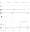
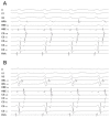
A narrow QRS complex Tachycardia with an apparently concentric retrograde atrial activation sequence
- PMID: 19308284
- PMCID: PMC2655075
A narrow QRS complex Tachycardia with an apparently concentric retrograde atrial activation sequence
Abstract
The retrograde atrial activation sequence constitutes an initial important clue to elucidate the tachycardia mechanism during diagnostic electrophysiological testing in patients with supraventricular tachycardia. However, in some cases its correct analysis is challenging.
Keywords: Supraventricular tachycardia; catheter ablation; mitral isthmus.
Figures


Comment in
-
Letter by Namboodiri regarding article, "a narrow QRS complex tachycardia with apparently concentric retrograde atrial activation sequence"--alternative mechanisms.Indian Pacing Electrophysiol J. 2009 Jul 1;9(4):211-2. Indian Pacing Electrophysiol J. 2009. PMID: 19652731 Free PMC article. No abstract available.
-
Letter by Mahajan regarding article, "a narrow QRS complex tachycardia with apparently concentric retrograde atrial activation sequence".Indian Pacing Electrophysiol J. 2009 Jul 1;9(4):213. Indian Pacing Electrophysiol J. 2009. PMID: 19652732 Free PMC article. No abstract available.
References
-
- Knight BP, et al. Diagnostic value of tachycardia features and pacing maneuvers during paroxysmal supraventricular tachycardia. J Am Coll Cardiol. 2000;36:574–582. - PubMed
-
- Michaud GF, et al. Differentiation of atypical atrioventricular node re-entrant tachycardia from orthodromic reciprocating tachycardia using a septal accessory pathway by the response to ventricular pacing. J Am Coll Cardiol. 2001;38:1163–1167. - PubMed
-
- Fiala M, et al. Mitral isthmus conduction block after a single radiofrequency application for a left concealed accessory pathway. J Cardiovasc Electrophysiol. 2007;18:1218–1219. - PubMed
-
- Luria DM, et al. Intra-atrial conduction block along the mitral valve annulus during accessory pathway ablation: evidence for a left atrial "isthmus". J Cardiovasc Electrophysiol. 2001;12:744–749. - PubMed
LinkOut - more resources
Full Text Sources
