

Depletion of serotonin and selective inhibition of 2B receptor suppressed tumor angiogenesis by inhibiting endothelial nitric oxide synthase and extracellular signal-regulated kinase 1/2 phosphorylation
- PMID: 19308295
- PMCID: PMC2657884
- DOI: 10.1593/neo.81630
Depletion of serotonin and selective inhibition of 2B receptor suppressed tumor angiogenesis by inhibiting endothelial nitric oxide synthase and extracellular signal-regulated kinase 1/2 phosphorylation
Abstract
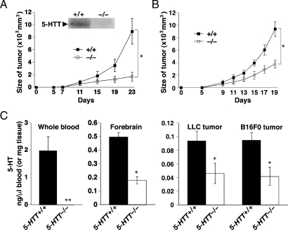
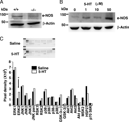
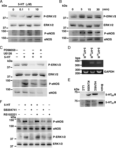
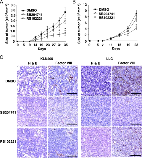
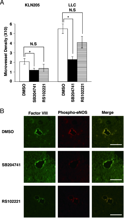
The effects of serotonin (5-HT) on tumor growth are inconsistent. We investigated whether a decreased level of 5-HT affected tumor growth using 5-HT transporter knockout (5-HTT(-/-)) mice, which showed 5-HT depletion. When cancer cells were injected subcutaneously into both 5-HTT(-/-) and 5-HTT(+/+) mice, the tumor growth was markedly attenuated in 5-HTT(-/-) mice. Serotonin levels in the blood, forebrain, and tumors of 5-HTT(-/-) mice bearing tumors were significantly smaller than those of their 5-HTT(+/+) littermates. However, 5-HT did not increase cancer cells' proliferation in vitro. When we applied 5-HTT inhibitors to the wild mice bearing tumors, they did not inhibit tumor growth. The endothelial nitric oxide synthase (eNOS) expressions in tumors were reduced in 5-HTT(-/-) mice compared with 5-HTT(+/+) mice. Stimulations with 5-HT (1-50 microM) induced eNOS expressions in human umbilical vein endothelial cell (HUVEC) in a concentration-dependent manner. When we measured activations of multiple signaling pathways by using a high-throughput phosphospecific antibodies platform, 5-HT stimulated the extracellular signal-regulated kinase 1/2 (ERK1/2) in HUVEC. Moreover, we found that the physiological level of 5-HT induced phosphorylation of both ERK1/2 and eNOS in HUVEC. Human umbilical vein endothelial cell expressed both 5-HT(2B) and 5-HT(2C) receptors. SB204741, a specific 5-HT(2B) receptor inhibitor, blocked 5-HT-induced ERK1/2 and eNOS phosphorylations, whereas RS102221, a specific 5-HT(2C) receptor inhibitor, did not in HUVEC. SB204741 reduced microvessel density in tumors and inhibited the proliferation of HUVEC in vitro. These results suggest that regulation of 5-HT and 5-HT receptors, especially the 5-HT(2B) receptor, may serve as a therapeutic strategy in cancer therapy.
Figures

Similar articles
-
Water extract of Korean red ginseng stimulates angiogenesis by activating the PI3K/Akt-dependent ERK1/2 and eNOS pathways in human umbilical vein endothelial cells.Biol Pharm Bull. 2007 Sep;30(9):1674-9. doi: 10.1248/bpb.30.1674. Biol Pharm Bull. 2007. PMID: 17827719
-
Endothelium-derived nitric oxide (NO) activates the NO-epidermal growth factor receptor-mediated signaling pathway in bradykinin-stimulated angiogenesis.Arch Biochem Biophys. 2014 Sep 15;558:14-27. doi: 10.1016/j.abb.2014.06.011. Epub 2014 Jun 21. Arch Biochem Biophys. 2014. PMID: 24960080
-
Glycogen synthase kinase-3β is critical for interferon-α-induced serotonin uptake in human Jurkat T cells.J Cell Physiol. 2012 Jun;227(6):2556-66. doi: 10.1002/jcp.22994. J Cell Physiol. 2012. PMID: 21898401
-
Skeletal effects of serotonin (5-hydroxytryptamine) transporter inhibition: evidence from in vitro and animal-based studies.J Musculoskelet Neuronal Interact. 2008 Apr-Jun;8(2):121-32. J Musculoskelet Neuronal Interact. 2008. PMID: 18622081 Free PMC article. Review.
-
Serotonin receptors and heart valve disease--it was meant 2B.Pharmacol Ther. 2011 Nov;132(2):146-57. doi: 10.1016/j.pharmthera.2011.03.008. Epub 2011 Apr 2. Pharmacol Ther. 2011. PMID: 21440001 Free PMC article. Review.
Cited by
-
Hippocampal angiogenesis and progenitor cell proliferation are increased with antidepressant use in major depression.Biol Psychiatry. 2012 Oct 1;72(7):562-71. doi: 10.1016/j.biopsych.2012.04.024. Epub 2012 May 30. Biol Psychiatry. 2012. PMID: 22652019 Free PMC article.
-
Transcription Profile and Pathway Analysis of the Endocannabinoid Receptor Inverse Agonist AM630 in the Core and Infiltrative Boundary of Human Glioblastoma Cells.Molecules. 2022 Mar 22;27(7):2049. doi: 10.3390/molecules27072049. Molecules. 2022. PMID: 35408449 Free PMC article.
-
Shaping the future of liver surgery: Implementation of experimental insights into liver regeneration.Eur Surg. 2018;50(3):132-136. doi: 10.1007/s10353-018-0515-3. Epub 2018 Mar 6. Eur Surg. 2018. PMID: 29875802 Free PMC article.
-
International Union of Basic and Clinical Pharmacology. CX. Classification of Receptors for 5-hydroxytryptamine; Pharmacology and Function.Pharmacol Rev. 2021 Jan;73(1):310-520. doi: 10.1124/pr.118.015552. Pharmacol Rev. 2021. PMID: 33370241 Free PMC article. Review.
-
The interconnectedness of cancer cell signaling.Neoplasia. 2011 Dec;13(12):1183-93. doi: 10.1593/neo.111746. Neoplasia. 2011. PMID: 22241964 Free PMC article.
References
-
- Nakaya N, Tsubono Y, Hosokawa T, Nishino Y, Ohkubo T, Hozawa A, Shibuya D, Fukudo S, Fukao A, Tsuji I, et al. Personality and the risk of cancer. J Natl Cancer Inst. 2003;95:799–805. - PubMed
-
- Asada M, Ebihara S, Okazaki T, Takahashi H, Yasuda H, Sasaki H. Tiapride may accelerate lung cancer in older people: a case report. J Am Geriatr Soc. 2005;53:731–732. - PubMed
-
- Asada M, Ebihara S, Numachi Y, Okazaki T, Yamanda S, Ikeda K, Yasuda H, Sora I, Arai H. Reduced tumor growth in a mouse model of schizophrenia, lacking the dopamine transporter. Int J Cancer. 2008;123:511–518. - PubMed
-
- Cattaneo MG, Codignola A, Vicentini LM, Clementi F, Sher E. Nicotine stimulates a serotonergic autocrine loop in human small-cell lung carcinoma. Cancer Res. 1993;53:5566–5568. - PubMed
-
- Cattaneo MG, Fesce R, Vicentini LM. Mitogenic effect of serotonin in human small cell lung carcinoma cells via both 5-HT1A and 5-HT1D receptors. Eur J Pharmacol. 1995;291:209–211. - PubMed
Publication types
MeSH terms
Substances
LinkOut - more resources
Full Text Sources
Molecular Biology Databases
Miscellaneous