Review
. 2009 Jul;66(14):2231-47.
doi: 10.1007/s00018-009-0014-6. Epub 2009 Mar 24.

Functional aspects of protein flexibility

Affiliations
Review

Functional aspects of protein flexibility

Kaare Teilum et al. Cell Mol Life Sci. 2009 Jul.

Abstract

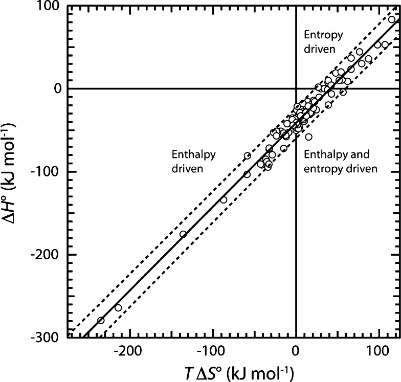
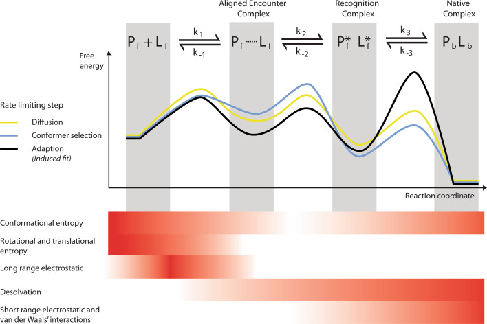
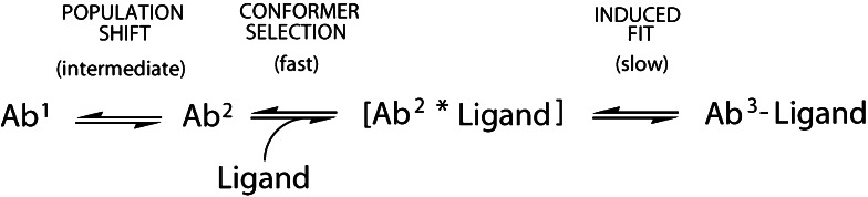
Proteins are dynamic entities, and they possess an inherent flexibility that allows them to function through molecular interactions within the cell, among cells and even between organisms. Appreciation of the non-static nature of proteins is emerging, but to describe and incorporate this into an intuitive perception of protein function is challenging. Flexibility is of overwhelming importance for protein function, and the changes in protein structure during interactions with binding partners can be dramatic. The present review addresses protein flexibility, focusing on protein-ligand interactions. The thermodynamics involved are reviewed, and examples of structure-function studies involving experimentally determined flexibility descriptions are presented. While much remains to be understood about protein flexibility, it is clear that it is encoded within their amino acid sequence and should be viewed as an integral part of their structure.


Figures

Fig. 1
Fig. 1
Schematic illustration of conformational events in proteins. The motions that change a protein’s conformation occur with time constants from nanoseconds to hours. The fastest motions, vibrations and librations around covalent bonds, result in atoms moving only a fraction of an Ångström. Ligand binding may involve only subtle motions like rearrangement of amino-acid residues in the binding site or larger movements over several Ångströms like domain reorientations as shown here for the binding of CR56 from LRP to RAP [123]. Similar movements may be observed for allosteric structural changes where binding of a ligand at one site of the protein changes the structure in another part. This is shown here for myosin V where binding and conversion of ATP to ADP in the upper domain (green) induces structural rearrangements not only around the nucleotide-binding site, but also in the distant lever arm domain (grey) [164, 165]. The ADP molecule is shown in colored spheres in the upper domain. Lastly, protein folding involves large movements of the whole protein chain as illustrated here for acyl-coenzyme A binding protein [166]. The fundamental terms dynamics and flexibility are defined at the bottom of the figure. Images of structures were generated in PyMol (DeLano Scientific)
Fig. 2
Fig. 2
Entropy–enthalpy compensation. Plot of ΔH° versus TΔS° for the binding equilibria of 100 different protein–protein or protein–peptide interactions. The solid line represents the best fit of a straight line to the data (slope = 1.00 ± 0.04). The dashed lines indicate the 95% prediction interval from the fit. Data from [–21, 167]
Fig. 3
Fig. 3
The flexible protein recognition model (FPRM). The protein-binding process is divided into three steps. First the two molecules in the unbound ensembles, Pf and Lf, encounter by diffusion to form the encounter complex [Pf⋯Lf] driven mostly by long-range forces. Within the encounter complex environment, a gradual desolvation is initiated, and while still populating the free ensembles, the two molecules form the recognition complex via conformer selection [Pf*Lf*] initiating a population shift. At this point short-range forces are in play. Within the recognition complex, induced fit and desolvation drive the formation of the final complex. Dependent on the barrier heights of the reactions, the protein-binding process may be controlled by diffusion (yellow line), by conformer selection (blue line) or by induced fit (large scale conformational changes) (black line). The forces that dominate the different steps during binding are shown schematically below. The intensity in color is relative between the different forces and not referring to any absolute magnitude. Formation of unaligned encounter complexes has been suggested, leading to a four-step binding reaction [168, 169]. Unaligned encounter complexes are not included in the FPRM described here. Figure modified from [69]
Fig. 4
Fig. 4
Large amplitude movements in enzyme activation. Two experimentally determined structures of a cysteine protease from Streptococcus pyogenes with the long latency and short switch loops highlighted in red. The dotted hand-drawn lines connect the backbone where electron density did not support model building. Left the crystal structure of the zymogen (1DKI, shown without the pro-domain for clarity) and right the NMR solution structure of the proteolytically activated monomer (2JTC). The latency loop moves >25 Å upon removal of the pro-domain and the protein folds up into an activated form
Fig. 5
Fig. 5
Binding of ligands to the Spe7 antibody. Two conformers, Ab1 and Ab2, are in equilibrium, and only the Ab2 conformer binds the ligand. The complex undergoes induced fit resulting in the stable Ab3-ligand complex. The relative rates of the individual reactions are indicated

References

    1. Dodson G, Verma CS. Protein flexibility: its role in structure and mechanism revealed by molecular simulations. Cell Mol Life Sci. 2006;63:207–219. doi: 10.1007/s00018-005-5236-7. - DOI - PMC - PubMed
    1. Dodson GG, Lane DP, Verma CS. Molecular simulations of protein dynamics: new windows on mechanisms in biology. EMBO Rep. 2008;9:144–150. doi: 10.1038/sj.embor.7401160. - DOI - PMC - PubMed
    1. Maragakis P, Lindorff-Larsen K, Eastwood MP, Dror RO, Klepeis JL, Arkin IT, Jensen MO, Xu H, Trbovic N, Friesner RA, Iii AG, Shaw DE. Microsecond molecular dynamics simulation shows effect of slow loop dynamics on backbone amide order parameters of proteins. J Phys Chem B. 2008;112:6155–6158. doi: 10.1021/jp077018h. - DOI - PMC - PubMed
    1. Linderstrøm-Lang K. Deuterium exchange between peptides and water. Chem Soc Spec Publ. 1955;2:1–20.
    1. Hvidt A. Deuterium exchange between ribonuclease and water. Biochim Biophys Acta. 1955;18:306–308. doi: 10.1016/0006-3002(55)90083-4. - DOI - PubMed

Publication types

LinkOut - more resources