

doi: 10.1007/s11832-007-0029-1.
Epub 2007 Jun 26.
The discoid meniscus
Affiliations
- PMID: 19308479
- PMCID: PMC2656711
- DOI: 10.1007/s11832-007-0029-1
Item in Clipboard
The discoid meniscus
J Child Orthop.
2007 Jul.
Abstract
Discoid lateral meniscus is an intra-articular knee disorder typically presented in the young population and during adolescence. Different types of meniscal disorders and varied forms of presentation have been reported. The natural history depends on the type of anomaly and the presence of symptoms. Management of the disorder should be directed toward the resolution of the symptoms while preserving meniscal tissue and function. Modern surgical techniques enable suturing and preservation of meniscal tissue. The clinical manifestations, diagnostic modalities and criteria, accompanying conditions and practical management considerations are reviewed.
Figures
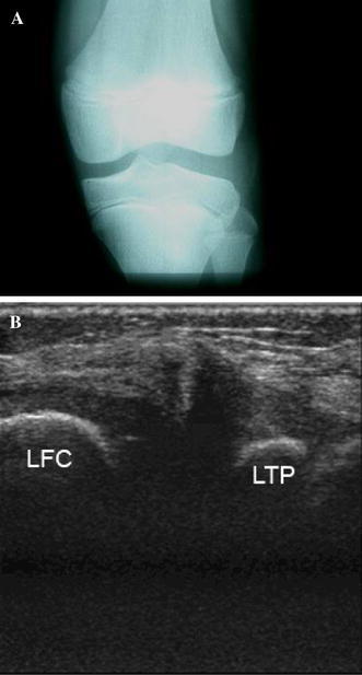
a Plain X-ray (anterior–posterior view) of a 10-year-old boy’s knee demonstrating widening of the lateral joint space, squaring of the lateral femoral condyle, cupping of the lateral tibial plateau, and tibial eminence hypoplasia. b An ultrasound of the wide and irregular lateral meniscus (LFC lateral femoral condyle, LTP lateral tibial plateau)
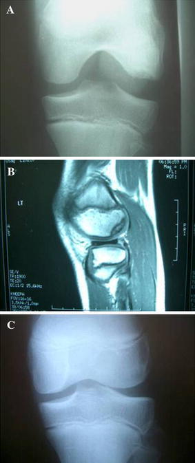
a Tunnel view of the left knee demonstrating signs of lateral discoid meniscus accompanied by an osteochondral lesion of the lateral femoral condyle. b Magnetic resonance imaging: a sagittal view demonstrating the same lesion. c Same knee 6 months after reshaping of the lateral discoid meniscus (note resolution of the osteochondral lesion)
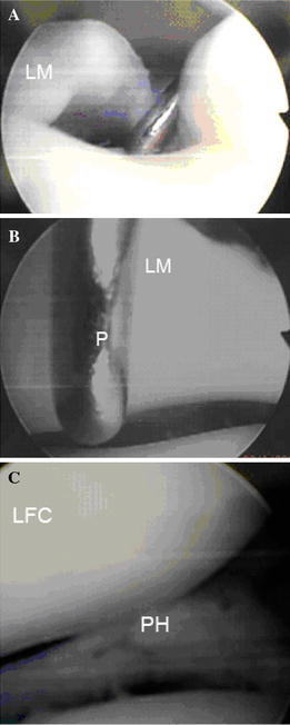
Arthroscopic demonstration of complete lateral discoid meniscus. a Wide medial edge of the lateral meniscus. The arthroscopic probe points the small tibial plateau part not covered with the meniscus (LM lateral meniscus). b Demonstration of the wideness architecture of the discoid meniscus inner edge, more than the 3-mm long arthroscopic probe (LM lateral meniscus, P Probe). c Same meniscus after reshaping and suturing of the posterior unstable horn (LFC lateral femoral condyle, PH posterior horn lateral meniscus, TP tibial plateau)
References
-
- Young R. The external semilunar cartilage as a complete disc. In: Cleland J, Mackay J, Young R, editors. Memoirs and memoranda in anatomy. London: Williams and Norgate; 1889. p. 179.
-
- Andrish J. Meniscal injuries in children and adolescents: diagnosis and management. J Am Acad Orthop Surg. 1996;4:231–237. - PubMed
-
- Kaplan EB. Discoid lateral meniscus of the knee joint. Bull Hosp Joint Dis. 1955;16:111–124. - PubMed
-
- Clark C, Ogden J. Development of the menisci of the human joint: morphologic changes and their potential role in childhood meniscal injury. J Bone Joint Surg Am. 1983;65:538–547. - PubMed
LinkOut - more resources
Full Text Sources
