. 2007 Mar;1(1):3-17.
doi: 10.1007/s11832-007-0006-8. Epub 2007 Feb 23.

Current approach to fibrous dysplasia of bone and McCune-Albright syndrome

Affiliations

Current approach to fibrous dysplasia of bone and McCune-Albright syndrome

Arabella I Leet et al. J Child Orthop. 2007 Mar.

Abstract

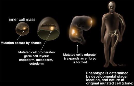
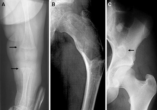
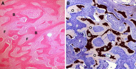
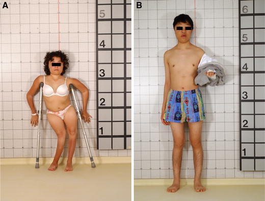
Fibrous dysplasia (FD) of bone is an uncommon disease caused by sporadic, congenital mutations in the cAMP regulating protein, G(s)alpha. It is an example of somatic mosaicism in which a wide spectrum of disease is possible. Widespread skeletal involvement is often associated with varying combinations of café-au-lait skin spots, and/or endocrine dysfunction (precocious puberty, renal phosphate wasting, hyperthyroidism, and/or growth hormone excess). Unrecognized and untreated endocrine dysfunction can exacerbate the skeletal disease. The diagnosis is usually established on clinical grounds on the basis of physical examination and typical radiographic appearance. Occasionally, gene testing of affected tissue may be helpful. The skeletal sites involved with disease are established at an early age, and the complications of fracture deformity are most pronounced in childhood. Bone pain in the absence of a fracture is more common in adults, but can also be present in children. Treatment with bisphosphonates is usually effective at relieving pain, but probably has no effect on the natural history of the disease. Scoliosis, which was previously thought to be an uncommon occurrence, has been shown to be common and progressive, and as such, warrants investigation and, when necessary, surgical treatment. The surgical management of FD remains challenging. Timing and technique remain controversial, but some consensus exists in that grafting materials (of any type) usually fail and should not be a central aspect of the surgical approach. Intramedullary devices are in general superior to side plates and screws. In extremely widespread disease with very early fracture and deformity, no surgical approach will affect final functional outcome. Efforts should be made for the initiation of international collaborative studies to better define optimal surgical approaches to the treatment of this challenging disease.


Figures

Fig. 1
Fig. 1
Development of fibrous dysplasia and McCune–Albright syndrome. The cartoon depicts the widely accepted hypothesis that FD/MAS is the result of somatic mutations that occur early in embryogenesis
Fig. 2
Fig. 2
Radiographic appearance of FD. a In the very young, the lesions can have a streaky appearance, and be found in association with rickets (arrows). b The typical radiographic appearance of FD is described as “ground glass.” As seen in this radiograph of the proximal femur, which also demonstrates the pathognomonic, shepherd’s crook. c FD in older patients, or patients with relatively inactive disease will demonstrate a sclerotic rim around the FD (arrow)
Fig. 3
Fig. 3
Histopathological appearance of FD. a H&E staining showing the classic “Chinese characters” made of trabeculae of bone (labeled B) with fibrous tissue interwoven between (labeled F). b Von Kossa stain for mineralization shows that the trabeculae of bone are composed of bone (black, labeled B) as well as osteoid (pale blue tissue labeled O). The fibrous tissue is labeled (F). The presence of the unmineralized bone confirms the osteomalacic nature of the lesions
Fig. 4
Fig. 4
The varied phenotype of two patients with McCune–Albright syndrome. The patient depicted in a has had premature closure of the physes from precocious puberty, and that, together with lower extremity bowing (windswept deformity), has resulted in short stature. The patient in b has acromegaly in addition to precocious puberty, thus the presence of growth hormone “rescued” him from short stature
Fig. 5
Fig. 5
CT for detecting subtle fractures. a This radiograph was from a 9-year-old boy who complained of new-onset, focal pain in the groin. No discernable fracture is apparent. bd Reformatted CT images of the lesion revealed a fracture in the medial proximal femur (arrows)
Fig. 6
Fig. 6
Use of CT imaging in FD. a This radiograph of a 10-year-old girl with FD revealed severe disease in both femurs, with a suggestion of a fracture. At the time of the radiograph, she was asymptomatic and ambulating without assistance. b CT at the level of the femora neck revealed the true extent of the disease, with almost complete disruption of the neck (arrows). The extent of the disease was not fully appreciated on the radiograph
Fig. 7
Fig. 7
Aneurysmal bone cyst in FD. Aneurysmal bone cysts are uncommonly associated with fibrous dysplasia. a The radiograph demonstrated disruption of the cortex. b MRI imaging revealed a clear-cut fluid-fluid level (arrows), consistent with an aneurysmal bone cyst. These lesions can be aggressive, eroding bone as shown by the loss of the cortex in a and b, and bleed extensively. Adequate blood should be obtained if surgical intervention is planned
Fig. 8
Fig. 8
Café-au-lait spots found in MAS. a This girl demonstrates typical skin lesions on the face with jagged “coast-of-Maine” borders. Note that skin lesions are bilateral, but that the lesion on the neck tends to respect the midline. b The lateral view of the same patient seen in a demonstrates how the lesions can sometimes tend to follow the developmental lines of Blashko. Also of note is the obvious goiter in this girl with hyperthyroidism. c Skin lesions can often be subtle. These lesions demonstrate coast-of-Maine borders and respect the midline. Again, note that there are lesions on both the right and left. The location of the lesion on the buttocks is a typical location and appearance for a skin lesion seen in association with FD. This girl had FD and skin spots, but no endocrine dysfunction
Fig. 9
Fig. 9
Scoliosis in association with FD. a The large magnitude curve in this 15-year-old girl, whose growth plates had been closed since the age of 10 due to precocious puberty, continued to progress after skeletal maturity. b A second patient who died from the complications of untreated scoliosis also developed this lesion after skeletal maturity. These dramatic cases emphasize the need to screen for, follow, and treat scoliosis in association with FD
Fig. 10
Fig. 10
Difficulty in managing proximal femur disease. a A proximal femoral head stress fracture that required stabilization is shown. b A hip screw and side plate plus bone grafting was used to treat the fracture. c A year later, the bone graft has resorbed and the plate, which ended in bone involved with FD, is starting to cause bending at the end of the plate

Similar articles

Cited by

References

    1. McCune DJ. Osteitis fibrosa cystica: the case of a nine-year-old girl who also exhibits precocious puberty, multiple pigmentation of the skin and hyperthyroidism. Am J Dis Child. 1936;52:743–744.
    1. Albright F, Butler AM, Hampton AO, Smith P. Syndrome characterized by osteitis fibrosa disseminata, areas, of pigmentation, and endocrine dysfunction, with precocious puberty in females: report of 5 cases. N Engl J Med. 1937;216:727–746. doi: 10.1056/NEJM193704292161701. - DOI
    1. Lichtenstein L. Polyostotic fibrous dysplasia. Arch Surg. 1938;36:874–898. doi: 10.1001/archsurg.1938.01190230153012. - DOI
    1. Lichtenstein L, Jaffe HL. Fibrous dysplasia of bone: a condition affecting one, several or many bones, graver cases of which may present abnormal pigmentation of skin, premature sexual development, hyperthyroidism or still other extraskeletal abnormalities. Arch Pathol. 1942;33:777–816.
    1. Dorfman HD, Czerniak B (1998) Fibroosseous lesions. In: Dorfman HD, Czerniak B (eds) Bone tumors. Mosby, St. Louis, pp 441–491

LinkOut - more resources