. 2008 Dec;2(6):407-15.
doi: 10.1007/s11832-008-0119-8. Epub 2008 Aug 29.

Lower-limb growth: how predictable are predictions?

Affiliations

Lower-limb growth: how predictable are predictions?

Paula M Kelly et al. J Child Orthop. 2008 Dec.

Abstract

Purpose: The purpose of this review is to clarify the different methods of predictions for growth of the lower limb and to propose a simplified method to calculate the final limb deficit and the correct timing of epiphysiodesis.

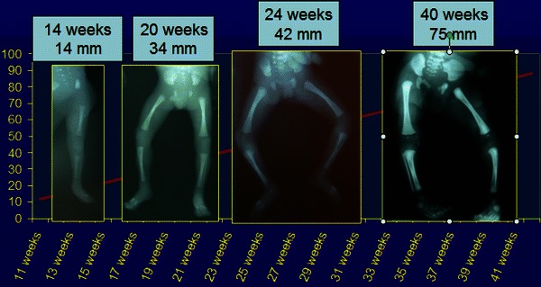
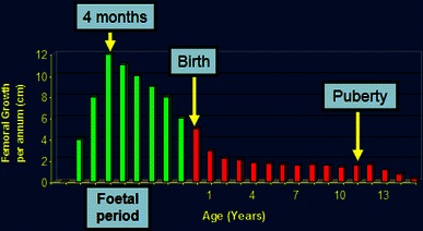
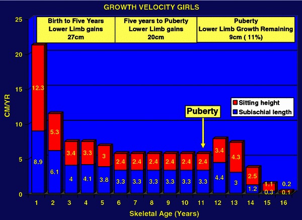
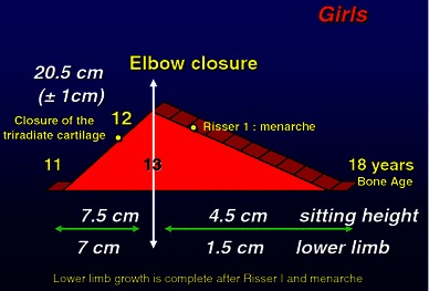
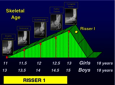
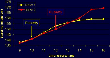
Background: Lower-limb growth is characterized by four different periods: antenatal growth (exponential); birth to 5 years (rapid growth); 5 years to puberty (stable growth); and puberty, which is the final growth spurt characterized by a rapid acceleration phase lasting 1 year followed by a more gradual deceleration phase lasting 1.5 years. The younger the child, the less precise is the prediction. Repeating measurements can increase the accuracy of predictions and those calculated at the beginning of puberty are the most accurate. The challenge is to reduce the margin of uncertainty. Confrontation of the different parameters-bone age, Tanner signs, annual growth velocity of the standing height, sub-ischial length and sitting height-is the most accurate method. Charts and diagrams are only models and templates. There are many mathematical equations in the literature; we must be able to step back from these rigid calculations because they are a false guarantee. The dynamic of growth needs a flexible approach. There are, however, some rules of thumb that may be helpful for different clinical scenarios.

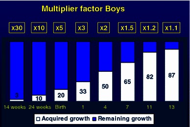
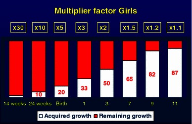
Calculation of limb length discrepancy: For congenital malformations, at birth the limb length discrepancy must be multiplied by 5 to give the final limb length discrepancy. Multiple by 3 at 1 year of age; by 2 at 3 years in girls and 4 years in boys; by 1.5 at 7 years in girls and boys, by 1.2 at 9 years in girls and 11 years in boys and by 1.1 at the onset of puberty (11 years bone age for girls and 13 years bone age for boys).

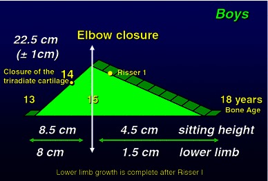
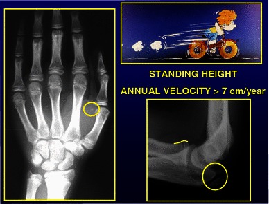
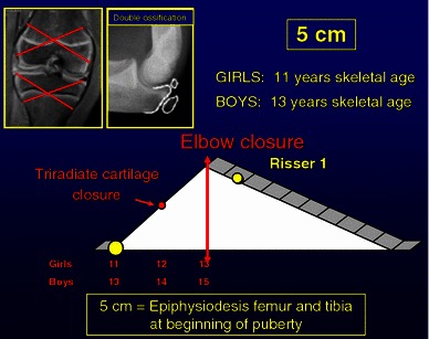
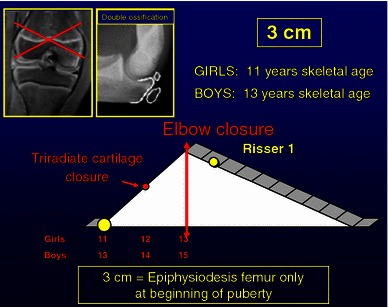
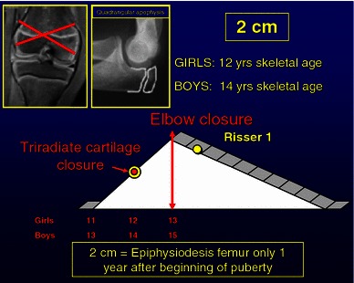
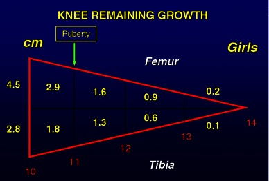
Timing of epiphysiodesis: For the timing of epiphysiodesis, several simple principles must be observed to reduce the margin of error; strict and repeated measurements, rigorous analysis of the data obtained, perfect evaluation of bone age with elbow plus hand radiographs and confirmation with Tanner signs. The decision should always be taken at the beginning of puberty. A simple rule is that, at the beginning of puberty, there is an average of 5 cm growth remaining at the knee. There are four common different scenarios: (1) A 5-cm discrepancy-epiphysiodesis of both femur and tibia at the beginning of puberty (11 years bone age girls and 13 years in boys). (2) A 4-cm discrepancy-epiphysiodesis of femur and tibia 6 months after the onset of puberty (11 years 6 months bone age girls, 13 years 6 months bone age boys, tri-radiate cartilage open). (3) A 3-cm discrepancy-epiphysiodesis of femur only at the start of puberty, (skeletal age of 11 years in girls and 13 years in boys). (4) A 2-cm discrepancy-epiphysiodesis of femur only, 1 year after the start of puberty (12 years bone age girls and 14 years in boys).


Figures

Fig. 1
Fig. 1
Foetal femoral diaphyseal growth with radiographic images at 14, 20, 24 and 40 weeks
Fig. 2
Fig. 2
Femoral growth velocity from the foetal period to skeletal maturity (girls) demonstrating a peak growth velocity at 4 months of antenatal life
Fig. 3
Fig. 3
Growth velocity in boys from birth to skeletal maturity
Fig. 4
Fig. 4
Growth velocity in girls from birth to skeletal maturity
Fig. 5
Fig. 5
Pubertal growth in boys [, p 44]
Fig. 6
Fig. 6
Pubertal growth in girls [, p 43]
Fig. 7
Fig. 7
Multiplier factor for boys from fetal life to skeletal maturity. At birth the acquired length is 20% therefore multiplier by 5 to calculate the estimated limb length discrepancy; multiply by 2 at 4 years of age and by 1.1 at the onset of puberty
Fig. 8
Fig. 8
Multiplier factor for girls from fetal life to skeletal maturity. At birth the acquired length is 20% therefore multiplier by 5 to calculate the estimated limb length discrepancy; multiply by 2 at 3 years of age and by 1.1 at the onset of puberty
Fig. 9
Fig. 9
Radiological changes at the onset of puberty, sesamoid ossification left hand, double ossification left olecranon apophysis
Fig. 10
Fig. 10
Pubertal diagram showing characteristic morphology of the left olecranon apophysis during puberty. Double ossification at onset of puberty, semilunar apophysis at puberty plus 6 months, quadrangular apophysis at puberty plus 1 year and complete fusion at peak pubertal growth velocity (puberty plus 2 years). (with permission Dimeglio et al., Accuracy of the sauvegrain method in determining skeletal age during puberty. JBJS (Am). 2005;87:1689–96.)
Fig. 11
Fig. 11
A 5-cm discrepancy can be corrected by performing an epiphysiodesis of both distal femur and proximal tibia at the beginning of puberty (11 years skeletal age girls, 13 years skeletal age boys).
Fig. 12
Fig. 12
A 4-cm discrepancy can be corrected by performing an epiphysiodesis of both distal femur and proximal tibia at puberty plus 6 months (11 years 6 months skeletal age girls, 13 years 6 months skeletal age boys)
Fig. 13
Fig. 13
A 3-cm discrepancy can be corrected by performing an epiphysiodesis of distal femur only at the beginning of puberty (11 years skeletal age girls, 13 years skeletal age boys)
Fig. 14
Fig. 14
A 2-cm discrepancy can be corrected by performing an epiphysiodesis of distal femur only at puberty plus 1 year (12 years skeletal age girls, 14 years skeletal age boys)
Fig. 15
Fig. 15
Knee growth (distal femur and proximal tibia) remaining in girls 10–14 years of age [, p 59]
Fig. 16
Fig. 16
Knee growth (distal femur and proximal tibia) remaining in boys 10–16 years of age [, p 59]
Fig. 17
Fig. 17
Growth chart of standing height for two sisters demonstrating the unpredictability of chronological age. Sister 1 commenced puberty at 10 years of age, sister 2 commenced at 12 years of age, with a resultant 10-cm difference in final height

Similar articles

Cited by

References

    1. Dimeglio A. La coissance en orthopedie. Montpellier: Sauramps; 1987.
    1. Dimeglio A. Growth in pediatric orthopaedics. J Pediatr Orthop. 2001;21(4):549–555. - PubMed
    1. Snijders RJ, Nicolaides K. Fetal biometry at 14–40 weeks gestation. Ultrasound Obstet Gynaecol. 1994;4(1):34–58. doi: 10.1046/j.1469-0705.1994.04010034.x. - DOI - PubMed
    1. Chitty LS, Altman DG, Henderson A, Campbell S. Charts of fetal size: 4. Femur length. Br J Obstet Gynaecol. 1994;101:132–135. doi: 10.1111/j.1471-0528.1994.tb13078.x. - DOI - PubMed
    1. Guihard-Costa AMTO, Droulle P, Larroche JC. Growth velocity of the biparietal diameter, abdominal diameter and femur length in the fetal period. Early Hum Dev. 1991;27:93–102. doi: 10.1016/0378-3782(91)90030-7. - DOI - PubMed