. 2009 Feb;3(1):11-20.
doi: 10.1007/s11832-008-0135-8. Epub 2008 Sep 27.

One-stage hip reconstruction in late neglected developmental dysplasia of the hip presenting in children above 8 years of age

Affiliations

One-stage hip reconstruction in late neglected developmental dysplasia of the hip presenting in children above 8 years of age

Hazem Mossad El-Tayeby. J Child Orthop. 2009 Feb.

Abstract

Purpose: To assess the clinical and radiological results of one-stage hip reconstruction for late neglected developmental dysplasia of the hip (DDH) in children above 8 years of age.

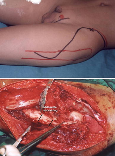
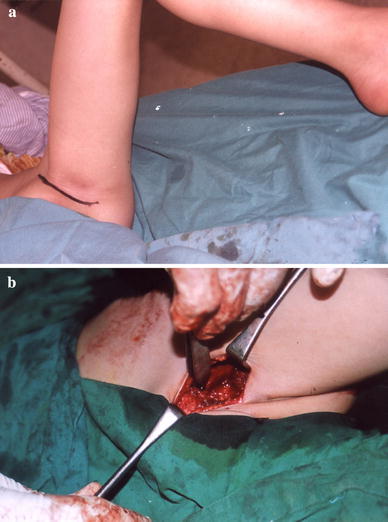
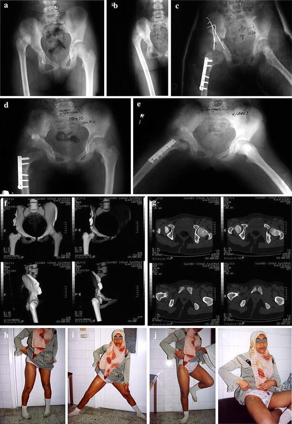
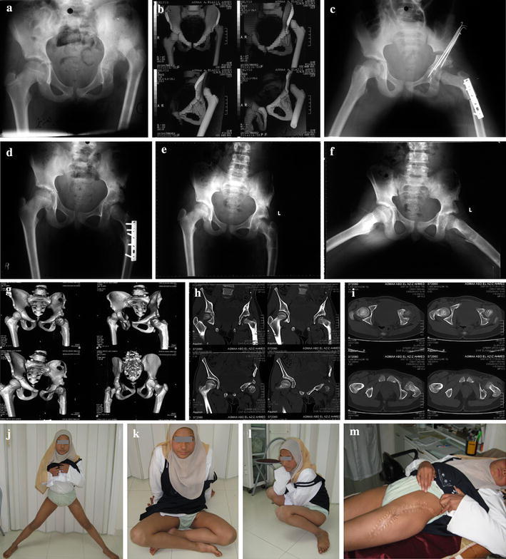
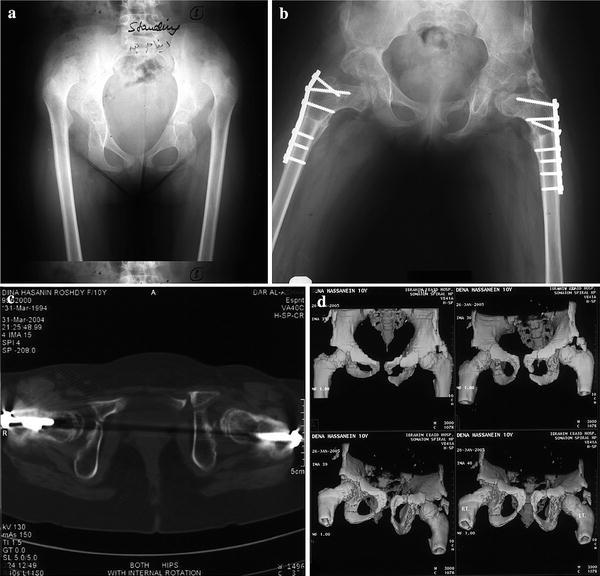
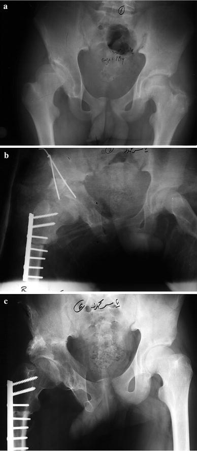
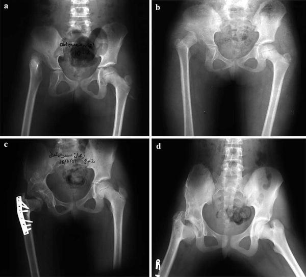
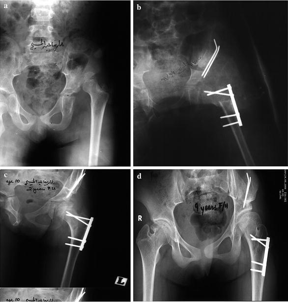
Methods: Nineteen hips in 16 patients, 14 females and two males (three being bilateral), were treated by open reduction adequate shortening (up to 5 cm) with derotation, and limited varization if needed, tight capsulorrhaphy, and appropriate pelvic reconstruction (Salter or triple acetabular osteotomy). The average age at operation 10.6 years (range 8-18 years). The period of follow up ranged from 3 to 9 years. A modified approach for the hip joint and upper femur was utilized, allowing better exposure. Pre- and post-operative plain radiography was performed for all cases and at follow up. Computed tomography (CT) or multislice CT with 3D reconstruction were carried out pre-operatively for recent cases and post-operatively for all, and were found to be helpful in providing a panorama of the dysplastic hip and in planning the required surgery and assessing the results.

Results: According to the McKay modified criteria, 15 hips (79%) were clinically excellent to good, while four hips (21%) were fair to poor. Radiographically, according to the Severin modified criteria, 16 hips (84%) were excellent to good and three hips (16%) were fair to poor. Limb length discrepancy ranged from 0.5 to 2.1 cm, as measured by CT scanograms. Complications were avascular necrosis (AVN) in an early case due to limited femoral shortening with resultant excessive stress over the femoral head and subluxation in another case.

Conclusion: A one-stage hip reconstruction for late neglected cases of DDH have achieved excellent results if adequate shortening with derotation is performed, together with appropriate acetabular reconstruction and tight capsulorrhaphy. Varization should not replace part or all of the required shortening, and should be added if required.


Figures

Fig. 1a, b
Fig. 1a, b
The modified iliofemoral approach. a Skin incision extending from the mid iliac crest to a mid inguinal point, then downwards and posteriorly to the trochanteric flare, to continue parallel to the femoral shaft. b Following full exposure, the iliac bone, the hip joint all round, and the upper third of the femoral shaft are well exposed
Fig. 2
Fig. 2
a Adductor incision: a straight incision extending from the adductor tubercle down to the ischial tuberosity. b Adductor tenotomy along the inferior pubic ramus down to bone. Osteotomy of the pubic rami could be done in triple pelvic osteotomy
Fig. 3a–k
Fig. 3a–k
Right developmental dysplasia of the hip (DDH) in an 11-year-old girl with excellent outcome. a, b Pre-operative radiograph in internal rotation revealing a normal neck shaft angle with a relatively small femoral head. c Post-operative reduction, well-contained head, triple pelvic osteotomy with the excised femoral segment used as a graft to maintain acetabular displacement. d, e Radiograph at 2 years follow up in full abduction with extended hips. f, g Multislice computed tomography (CT) sections and 3D reconstruction revealing normal anteversion angle and a normal hip articulation. hk Photographs of the patient at 17 years of age with a mobile hip performing all movements
Fig. 4a–m
Fig. 4a–m
Left DDH in a 12-year-old girl with excellent outcome. a, b Pre-operative radiograph and 3D reconstruction CT views showing a highly dislocated relatively small femoral head. c Post-operative radiograph, with well-contained femoral head following 5 cm femoral shortening with derotation, minimal varization Salter pelvic osteotomy. d Two years follow up radiographs. e, f Three years follow up radiographs with full abduction in extension. gi Three years follow up CT with reconstruction views showing well-contained femoral head with normal anteversion angle. jm Photographs showing full range of movements of the left hip, yet with an unsightly scar
Fig. 5a–d
Fig. 5a–d
Bilateral DDH in a 10-year-old girl with good outcome. a Pre-operative radiograph showing highly dislocated femoral heads. b Late post-operative radiograph for both hips. The left was reconstructed first; a triple pelvic osteotomy was performed. The right side followed; a Salter osteotomy was found to be enough to cover the head. c, d CT with 3D reconstruction showing an excellent reconstruction, with accepted anteversion angle, yet the left hip had limitation of abduction
Fig. 6a–c
Fig. 6a–c
Right DDH in a 18-year-old male with good outcome. a Pre-operative radiograph showing a dysplastic acetabulum. b Post-operative radiograph. The head is well-contained and 3 cm shortening was needed to bring the head down and to compensate for the pelvic procedure. c Late follow up radiograph
Fig. 7a–d
Fig. 7a–d
Right DDH in a 9-year-old girl with a poor result. a, b Pre-operative radiographs in neutral and internal rotation. c Late post-operative view showing excessive varization at the expense of shortening with evident avascular necrosis (AVN). d Coxa vara 7 years later (aged 16 years)
Fig. 8a–d
Fig. 8a–d
Left DDH in an 8-year-old girl with a fair outcome. a Pre-operative radiograph. b Post-operative radiograph showing a well-contained femoral head with acceptable neck shaft angle. c Two years follow up radiograph showing coxa valga, yet still a well-contained head and intact Shenton line. d Eight years follow up (aged 16 years) showing severe coxa valga with subluxation

References

    1. Ashley RK, Larsen LJ, James PM. Reduction of dislocation of the hip in older children: a preliminary report. J Bone Joint Surg Am. 1972;54:545–550. - PubMed
    1. Berkeley ME, Dickson JH, Cain TE, Donovan MM. Surgical therapy for congenital dislocation of the hip in patients who are twelve to thirty-six months old. J Bone Joint Surg Am. 1984;66:412–420. - PubMed
    1. Coleman SS. Congenital dysplasia and dislocation of the hip. St. Louis, MO: CV Mosby; 1978. pp. 95–154.
    1. Lindstrom JR, Ponseti IV, Wenger DR. Acetabular development after reduction in congenital dislocation of the hip. J Bone Joint Surg Am. 1979;61:112–118. - PubMed
    1. Shih CH, Shih HN. One-stage combined operation of congenital dislocation of the hips in older children. J Pediatr Orthop. 1988;8:535–539. doi: 10.1097/01241398-198809000-00007. - DOI - PubMed