. 2009 Sep;5(3):335-42.
doi: 10.1007/s11302-009-9158-y. Epub 2009 Mar 24.

Vascular smooth muscle cell expression of ectonucleotidase CD39 (ENTPD1) is required for neointimal formation in mice

Affiliations

Vascular smooth muscle cell expression of ectonucleotidase CD39 (ENTPD1) is required for neointimal formation in mice

Amir Behdad et al. Purinergic Signal. 2009 Sep.

Abstract

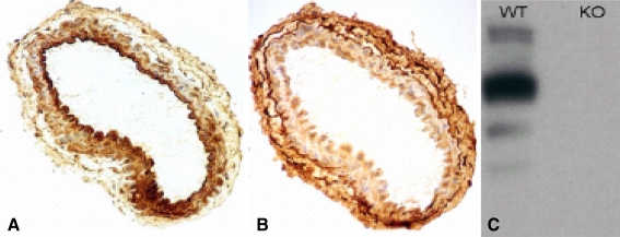
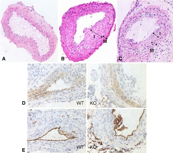
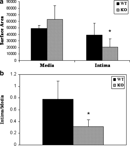
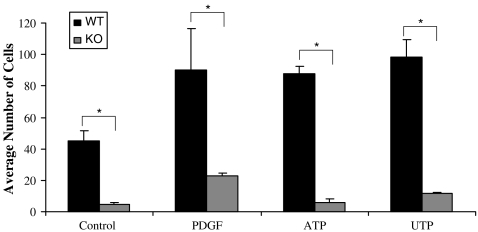
Vascular smooth muscle cell (VSMC) migration and proliferation are critical steps in the pathogenesis of atherosclerosis, post-angioplasty restenosis, neointimal hyperplasia, and chronic allograft rejection. Extracellular nucleotides are known to influence both migration and proliferation of VSMC. Although it is well established that vascular endothelial Cd39/ENTPD1 regulates blood nucleotide concentrations, whether Cd39 associated with VSMC also impacts vascular wall pathology has not been investigated. The objective of this paper is to determine levels of expression of Cd39 on VSMC and functional consequences of gene deletion in vitro and in vivo. Cd39 is the major ectonucleotidase in VSMC, as shown by substantive decreases in ecto-ATPase and -ADPase activity in Cd39-null cells compared to wild type. Significant decreases in neointimal lesion formation are observed in Cd39-null mice at 21 days post arterial balloon injury. Stimulated Cd39-null VSMC have pronounced proliferative responses in vitro. However, using Transwell systems, we show that Cd39-null VSMC fail to migrate in response to ATP, UTP, and PDGF. Cd39 is the dominant ectonucleotidase expressed by VSMC. Deletion of Cd39 in mice results in decreased neointimal formation after vascular injury and is associated with impaired VSMC migration responses in vitro.


Figures

Fig. 1
Fig. 1
ab Immunostaining of mouse carotid artery with anti-murine NTPDase1 and NTPDase2 polyclonal antibodies. a NTPDase 1 (Cd39) is predominantly expressed on the endothelial and the intimal surface. b In contrast, NTPDase 2 (Cd39L1) is mostly expressed on the adventitia layer of the vessels. c Western blotting of VSMC shows that Cd39 is expressed by the wild-type (WT) VSMC and is clearly absent in Cd39-null (knock-out) VSMC
Fig. 2
Fig. 2
Biochemical activity of intact murine VSMC (expressed as nanomole per minute per milligram). NTPDase activities of intact cells were determined by measurement of phosphate release from the respective substrates. Data are shown as for specific ATPase and ADPase activities in wild type (WT) and Cd39-null VSMC (mean ± SD). Cd39 is the dominant ectonucleotidase in VSMC and its deletion in null cells significantly decreases ATPase and ADPase activity (*P < 0.05)
Fig. 3
Fig. 3
Histopathologic features of injured common carotid artery in wild type and Cd39-null mice. ac H&E staining of control: WT-uninjured (KO-uninjured had similar features and is not shown here), WT- and Cd39 KO-injured carotid arteries (CA) 3 week after injury: KO vessels show less neointimal formation (I intima, M media). de Immunostaining of injured vessels with a smooth muscle actin monoclonal Ab. (d) and with CD31 monoclonal Ab to the endothelial marker (e) confirm that neointimal layer contains VSMC cells
Fig. 4
Fig. 4
Quantitative histomorphometry measurement post-wire injury of common carotid artery in wild type and Cd39-null mice. Twenty-one days after arterial injury, carotid arteries were collected from six animals of each group (WT and KO) and snap-frozen in liquid nitrogen. Samples were sectioned (5 µm) at 200 µm intervals, and stained with hematoxylin–eosin. Six images per vessel were acquired at various magnification levels. Distances and areas of intimal and medial layer were calculated by digital imaging software (Image J, NIH) using arbitrary parameters. a Intimal (I) and media (M) surface area compared in WT (n = 6) and KO (n = 6) carotid arteries. KO intimal surface area is significantly less than WT (*P < 0.05). There was no statistically significant difference between WT and KO media surface area (P = 0.09). b Neointimal formation shown by I/M ratio was shown to be significantly decreased in Cd39 null mice at day 21 following injury (*P < 0.05)
Fig. 5
Fig. 5
Proliferation assay in wild type and Cd39-null VSMC a: Proliferative response (thymidine incorporation) to serum and PDGF stimulation. Primary mouse VSMCs from control and Cd39-null mice were seeded on 96-well plates (1 × 104 cells/well) and subsequently serum-starved (0.2% FCS) for the following 24 h and then stimulated with 10% FCS containing medium, 50 nM PDGF for 24 h. Twelve hours prior to harvest, cells were pulsed with [3H]-thymidine. Columns, average of thymidine incorporated count ratio to controls, for three experiments (in triplicate for each experiment), bars, SE. *P < 0.05. b Proliferative response to different concentrations of ATP. Conditions are similar to prior study, except pulse stimulation with ATP was for 5 min; cells were kept in 0.2% FCS for completion of 24 h. Columns, average of thymidine incorporated count ratio to controls, for three experiments (in triplicate for each experiment), bars SE. *P < 0.05
Fig. 6
Fig. 6
Impaired migration of Cd39-null VSMC. Transwell systems were used to assess the chemotactic effects of PDGF, ATP, and UTP on cultured WT and Cd39-null VSMC. In these experiments, 5 × 106 cells were placed in each well. In the lower chamber, the medium consisted of basal media with 0.5% serum (used as control), and basal media with PDGF, ATP, and UTP (see text). Cells migrating to the lower surface of the filter within 6 h were counted. Columns show averages of cells for three experiments (in triplicate for each experiment), counted per 5 HPF/well, and indicating migratory responses; bars denote SE. *P < 0.005

References

    1. {'text': '', 'ref_index': 1, 'ids': [{'type': 'PubMed', 'value': '8594927', 'is_inner': True, 'url': 'https://pubmed.ncbi.nlm.nih.gov/8594927/'}]}
    2. Ellsworth ML, Forrester T, Ellis CG, Dietrich HH (1995) The erythrocyte as a regulator of vascular tone. Am J Physiol 269:H2155–H2161 - PubMed
    1. {'text': '', 'ref_index': 1, 'ids': [{'type': 'DOI', 'value': '10.1074/jbc.273.11.6334', 'is_inner': False, 'url': 'https://doi.org/10.1074/jbc.273.11.6334'}, {'type': 'PubMed', 'value': '9497362', 'is_inner': True, 'url': 'https://pubmed.ncbi.nlm.nih.gov/9497362/'}]}
    2. Hamada K, Takuwa N, Yokoyama K, Takuwa Y (1998) Stretch activates Jun N-terminal kinase/stress-activated protein kinase in vascular smooth muscle cells through mechanisms involving autocrine ATP stimulation of purinoceptors. J Biol Chem 273:6334–6340. doi:10.1074/jbc.273.11.6334 - PubMed
    1. {'text': '', 'ref_index': 1, 'ids': [{'type': 'DOI', 'value': '10.1074/jbc.272.39.24348', 'is_inner': False, 'url': 'https://doi.org/10.1074/jbc.272.39.24348'}, {'type': 'PubMed', 'value': '9305892', 'is_inner': True, 'url': 'https://pubmed.ncbi.nlm.nih.gov/9305892/'}]}
    2. Lazarowski ER, Homolya L, Boucher RC, Harden TK (1997) Direct demonstration of mechanically induced release of cellular UTP and its implication for uridine nucleotide receptor activation. J Biol Chem 272:24348–24354. doi:10.1074/jbc.272.39.24348 - PubMed
    1. {'text': '', 'ref_index': 1, 'ids': [{'type': 'PubMed', 'value': '2548757', 'is_inner': True, 'url': 'https://pubmed.ncbi.nlm.nih.gov/2548757/'}]}
    2. Coad SB, Pearson JD (1989) Metabolism of adenine nucleotides in human blood. Circ Res 65:531–537 - PubMed
    1. {'text': '', 'ref_index': 1, 'ids': [{'type': 'DOI', 'value': '10.1007/BF01741386', 'is_inner': False, 'url': 'https://doi.org/10.1007/bf01741386'}, {'type': 'PubMed', 'value': '2651791', 'is_inner': True, 'url': 'https://pubmed.ncbi.nlm.nih.gov/2651791/'}]}
    2. Luthje J (1997) Origin, metabolism and function of extracellular adenine nucleotides in the blood. Klin Wochenschr 67:317–327. doi:10.1007/BF01741386 - PubMed