. 2009 Dec;2(1):33-47.
doi: 10.1007/s12307-009-0019-6. Epub 2009 Mar 18.

Dual FGF-2 and intergrin alpha5beta1 signaling mediate GRAF-induced RhoA inactivation in a model of breast cancer dormancy

Affiliations

Dual FGF-2 and intergrin alpha5beta1 signaling mediate GRAF-induced RhoA inactivation in a model of breast cancer dormancy

Judith Barrios et al. Cancer Microenviron. 2009 Dec.

Abstract

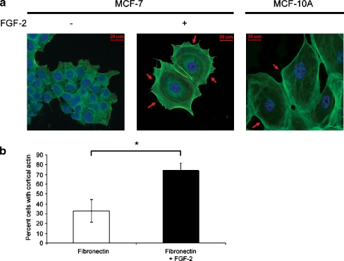
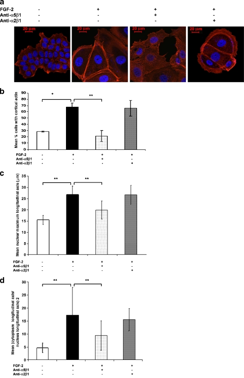
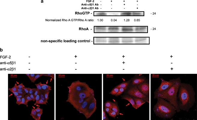
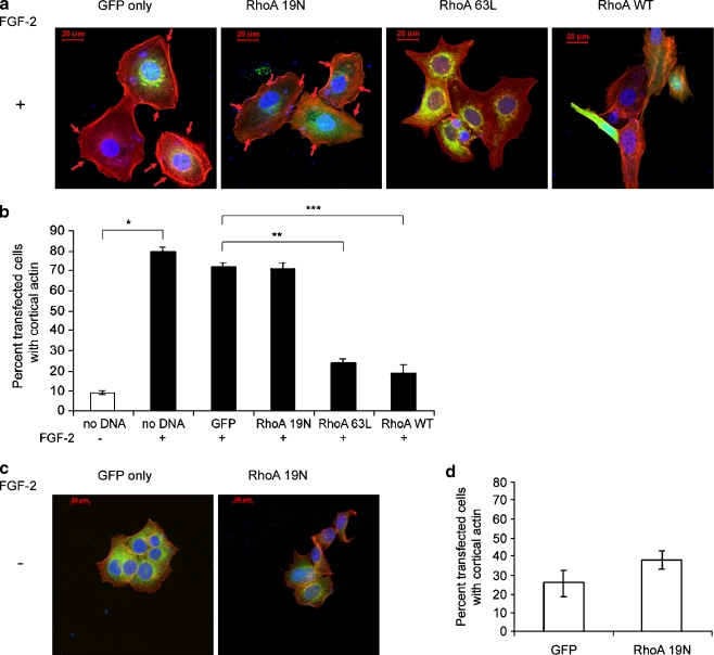
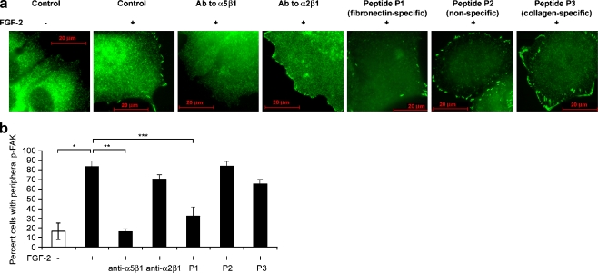
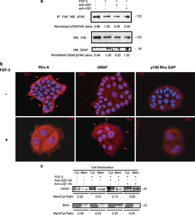
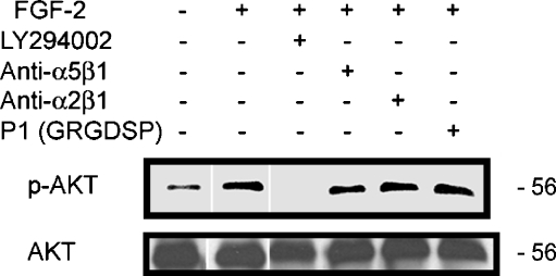
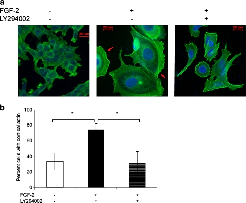
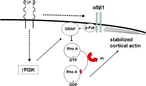
Interactions with the bone marrow stroma regulate dormancy and survival of breast cancer micrometastases. In an in vitro model of dormancy in the bone marrow, we previously demonstrated that estrogen-dependent breast cancer cells are partially re-differentiated by FGF-2, re-express integrin alpha5beta1 lost with malignant transformation and acquire an activated PI3K/Akt pathway. Ligation of integrin alpha5beta1 by fibronectin and activation of the PI3K pathway both contribute to survival of these dormant cells. Here, we investigated mechanisms responsible for the dormant phenotype. Experiments demonstrate that integrin alpha5beta1 controls de novo cytoskeletal rearrangements, cell spreading, focal adhesion kinase rearrangement to the cell perimeter and recruitment of a RhoA GAP known as GRAF. This results in the inactivation of RhoA, an effect which is necessary for the stabilization of cortical actin. Experiments also demonstrate that activation of the PI3K pathway by FGF-2 is independent of integrin alpha5beta1 and is also required for cortical actin reorganization, GRAF membrane relocalization and RhoA inactivation. These data suggest that GRAF-mediated RhoA inactivation and consequent phenotypic changes of dormancy depend on dual signaling by FGF-2-initiated PI3K activation and through ligation of integrin alpha5beta1 by fibronectin.


Figures

Fig. 1
Fig. 1
Cortical actin stabilization in dormant breast cancer cells. a MCF-7 cells incubated with or without FGF-2 10 ng/ml on fibronectin-coated cover slips for 6 days at clonogenic density were stained with BODIPY-Phallacidin (green actin staining) and DAPI (blue nuclear staining) and photographed at 400 x magnification (see Materials and Methods). A 20 μM size bar is included in all fluorescence photographs in all figures. MCF-10A cells were incubated on fibronectin-coated slides and stained in a similar manner as controls and demonstrate morphological similarity with dormant MCF-7 cells. Arrows indicate prominent places of cortical actin redistribution around the perimeter of the cytoplasm. b The percentages of cells with cortical actin distribution of >50% of the perimeter were manually counted in approximately 100 cells and graphed. Experiments were done in triplicate slides. Error bars are + standard deviations. *p < 0.01 (Student’s t test)
Fig. 2
Fig. 2
Cortical actin stabilization in dormant breast cancer cells is integrin α5β1-dependent. MCF-7 cells were incubated with or without FGF-2 10 ng/ml on fibronectin-coated cover slips at clonogenic density. Blocking antibodies to integrin α5β1 and integrin α2β1 2 μg/ml were added on day 3. Cells were stained with rhodamine phalloidin (red actin staining) and DAPI (blue nuclear staining) and photographed at 400 x magnification. A 20 μM size bar is included in all photographs. a Blocking antibody to integrin α5β1, the fibronectin receptor upregulated in dormant MCF-7 cells, reversed the cortical redistribution of fibrillar actin, the increased nuclear size and the increased cytoplasm to nucleus ratios in dormant cells. Blocking antibody to integrin α2β1, the upregulated collagen receptor, had no effect. b Quantitative representation of the percentage of manually counted cells with cortical actin on triplicate slides from a duplicate experiment. c Quantitative representation of the maximal longitudinal axis of the nuclei and d Quantitative representation of the square of the ratio of the maximal cytoplasm long axis to that of the maximal nuclear long axis. Error bars are + standard deviations. *p < 0.005, **p < 0.001 (Student’s t test)
Fig. 3
Fig. 3
Downregulation of RhoA GTP-loading in dormant MCF-7 breast cancer cells is integrin α5β1-dependent. a Cells were incubated on 10–12 fibronectin-coated 10 cm plates per condition and cultured as described. On day 6, equal cell numbers were lysed, incubated with Rhotekin-conjugated agarose, precipitated and analyzed by western blot with anti-RhoA antibody. Total lysates were used for RhoA western blot controls and a nonspecific band on the Coomasie-stained membrane was used as a loading control to confirm the increased protein content of dormant cells. RhoA GTP and total Rho bands were quantitated with a densitometer and ratios were normalized to that of growing cells. The experiment was done three times. b The RhoA GTP-loading data was corroborated by indirect immunofluorescence-staining of cells on fibronectin-coated cover slips with anti-RhoA antibody (red) and photography at 630 x magnification. Growing cells exhibited membrane localization of RhoA (arrows) which disappeared in dormant cells. Blocking antibody to integrin α5β1 2 μg/ml induced re-localization of RhoA to the membrane, while blocking antibody to integrin α2β1 2 μg/ml had only a minimal effect. Nuclear DAPI staining is shown in blue
Fig. 4
Fig. 4
Downregulation of RhoA GTP-loading is necessary but not sufficient for cortical actin rearrangement in dormant cells. Cells on fibronectin-coated cover slips in medium containing FGF-2 10 ng/ml (A. and B.) or lacking FGF-2 (C. and D.) were transiently transfected with 10:1 ratios of the three RhoA vectors and the GFP vector or with the GFP vector alone and stained with rhodamine phalloidin (red) and DAPI (blue nuclear staining). Cortical actin was identified and quantitated in the GFP-transfected green cells only. a Cortical distribution of F-actin was observed in GFP only- and RhoA 19N (dominant negative)-transfected dormant cells (arrows), but was markedly diminished in dormant cells transfected with RhoA63L (constitutively active) or RhoA wild type (RhoAWT). These latter two transfectants also induced the appearance of stress fibers. Cells were photographed at 400 x magnification. b Quantitative assessment of the percentage of cells with >50% cortical distribution demonstrates a statistically significant increase in cortical actin in dormant cells compared with growing cells (*p < 0.01), between GFP- and RhoA63L-transfected dormant cells (**p < 0.001) and between GFP- and RhoAWT-transfected dormant cells (***p < 0.02) (Student’s t test). Error bars are + standard deviations. All other differences were not statistically significant. c Transfection of growing cells with dominant negative RhoA19N did not induce either the dormant phenotype or actin rearrangement. Transfection with either constitutively active RhoA63L or wild type RhoA also did not affect cortical actin (not shown). D. Statistical comparison of cell distributions with cortical actin was not affected in growing cells by dominant negative RhoA19N, nor by the other vectors (not shown)
Fig. 5
Fig. 5
Peripheral phospho-Y397 FAK localization in dormant cells is integrin α5β1-dependent. a MCF-7 cells were incubated on fibronectin-coated cover slips with medium containing FGF-2 10 ng/ml. Blocking antibodies to integrin α5β1 and integrin α2β1 2 μg/ml and blocking peptides to fibronectin (P1), to collagen (P3), and a non-binding control (P2) 100 nM were added on day 3 as described in Materials and Methods. Cells were stained with antibodies to phospho-Y397 FAK on day 6 and photographed at 1,000 x magnification. Localization of phospho-Y397 FAK with dormancy is reversed by blocking fibronectin binding with blocking antibody to integrin α5β1 or blocking peptide P1 to fibronectin. b Graphic depiction of induction of peripheral phospho-Y397 FAK in dormant cells (*p < 0.005), and reversal of localization by blocking antibody to integrin α5β1 (**p < 0.001) and blocking peptide to fibronectin P1 (***p < 0.01) (Student’s t test). Error bars are + standard deviations. All other differences were not statistically significant. Data is from one of two duplicate experiments with triplicate slides with approximately 100 cells counted per slide
Fig. 6
Fig. 6
Integrin α5β1-dependent peripherally localized phospho-Y397 FAK in dormant cells is associated with membrane localization of the RhoA GAP GRAF. a Cells incubated on fibronectin-coated tissue culture plates with and without FGF-2 10 ng/ml with control or blocking antibodies to integrin α5β1 and integrin α2β1 2 μg/ml added on day 3 were harvested on day 6. Lysates from equal cell numbers were immunoprecipitated with antibody to FAK and stained on western blot with anti-phospho-Y397 FAK antibody, total FAK antibody and GRAF antibody. Bands were quantitated using a densitometer and ratios of phospho-FAK to FAK and GRAF to phospho-FAK were normalized to the bands from dormant cells in the immunoprecipitates. Ratios of phospho-FAK to total FAK and total FAK to control bands were also normalized to dormant cells. b GRAF membrane localization in dormant cells and the corresponding RhoA departure form its membrane localization was demonstrated on immunofluorescence-stained cells on fibronectin-coated cover slips (red) and photography at 630 x magnification. Growing cells exhibited membrane localization of RhoA (arrows) which disappeared in dormant cells, while GRAF membrane localization appeared in dormant cells (arrows). Immunostaining with antibody to p190 Rho GAP was used as a negative control, demonstrating no evident staining in either growing or dormant cells. Nuclear DAPI staining is shown in blue. c Membrane fractionation of growing and dormant cells with and without added blocking antibodies to integrin α5β1 and integrin α2β1 2 μg/ml and western blotting of isolates with antibody to GRAF and BAX, used as a cytoplasm-localizing control. Bands were quantitated using a densitometer and ratios of membrane- to cytoplasm-localizing GRAF and BAX were calculated
Fig. 7
Fig. 7
Akt activation by FGF-2 in dormant cells is independent of integrin α5β1 ligation. Western blots of lysates from cells incubated on fibronectin with and without FGF-2 10 ng/ml or blocking antibodies to integrin α5β1 or integrin α2β1 2 μg/ml, blocking peptide P1 to fibronectin 100 nm, or PI3K inhibitor LY294002 25 μM on day 3, as described in Materials and Methods, were stained with antibody to phospho-Akt or total Akt
Fig. 8
Fig. 8
Cortical actin stabilization in dormant breast cancer cells is PI3K-dependent. a MCF-7 cells incubated with or without FGF-2 10 ng/ml on fibronectin-coated cover slips at clonogenic density, with and without addition of LY294002 25 μM on day 3 were stained on day 6 with BODIPY-Phallacidin (green actin staining) and DAPI (blue nuclear staining) and photographed at 400 x magnification. The figure demonstrates cortical actin distribution that appears in dormancy and is reversed by PI3K inhibition. The appearance of stress fibers and loss of the characteristic cell spreading is evident in dormant cells inhibited by LY294002. b Quantitative representation of manually counted cells with cortical actin on triplicate slides from a duplicate experiment demonstrating an increase in cortical actin with dormancy and reversal with PI3K inhibition. Error bars are + standard deviations. *p < 0.01 (Student’s t test)
Fig. 9
Fig. 9
Membrane localization of GRAF in dormant cells is PI3K-dependent. a GRAF membrane localization in dormant cells and the corresponding RhoA departure form its membrane localization was demonstrated on immunofluorescence-stained cells on fibronectin-coated cover slips (red) and photography at 630 x magnification. Addition of LY294002 25 μM on day 3 to the incubation medium resulted in abrogation of the membrane localization of GRAF and a corresponding membrane re-localization of RhoA (arrows). Growing cells exhibited membrane localization of RhoA (arrows) which disappeared in dormant cells, while GRAF membrane localization appeared in dormant cells (arrows). Nuclear DAPI staining is shown in blue. b Membrane fractionation of growing and dormant cells with and without added LY294002 25 μM and western blotting of isolates with antibody to GRAF and BAX, used as a cytoplasm-localizing control, demonstrates that the membrane localization of GRAF in dormant cells is reversed by blocking of PI3K signaling. Bands were quantitated using a densitometer and ratios of membrane- to cytoplasm-localizing GRAF and BAX were calculated
Fig. 10
Fig. 10
Schema of dual FGFR and integrin α5β1 parallel steady state signaling in the dormancy model. The schema indicates FGF-2-initiated upregulation of integrin α5β1 which reaches steady state after several days. Dual signaling through FGFR through PI3K and independently through integrin α5β1 induces activation of FAK and membrane localization and activation of the RhoA GAP GRAF. This results in inactivation of RhoA and a permissive steady state for cortical rearrangement of F-actin

References

    1. Braun S, Pantel K, Muller P, et al. Cytokeratin-positive cells in the bone marrow and survival of patients with stage I, II, or III breast cancer. N Engl J Med. 2000;342:525–533. doi: 10.1056/NEJM200002243420801. - DOI - PubMed
    1. Braun S, Kentenich C, Janni W, et al. Lack of an effect of adjuvant chemotherapy on the elimination of single dormant tumor cells in bone marrow of high risk breast cancer patients. J Clin Onc. 2000;18:80–86. - PubMed
    1. Korah R, Boots M, Wieder R. Integrin α5β1 promotes survival of breast cancer cells: an in vitro paradigm for breast cancer cell dormancy in the bone marrow. Can Res. 2004;64:4514–4522. doi: 10.1158/0008-5472.CAN-03-3853. - DOI - PubMed
    1. Nguyen PL, Taghian AG, Katz MS, et al. Breast cancer subtype approximated by estrogen receptor, progesterone receptor, and HER-2 is associated with local and distant recurrence after breast-conserving therapy. J Clin Oncol. 2008;26:2373–2378. doi: 10.1200/JCO.2007.14.4287. - DOI - PubMed
    1. Haffty BG, Yang Q, Reiss M, et al. Locoregional relapse and distant metastasis in conservatively managed triple negative early-stage breast cancer. J Clin Oncol. 2006;24:5652–5657. doi: 10.1200/JCO.2006.06.5664. - DOI - PubMed

LinkOut - more resources