

Genotype-dependent tumor regression in Marek's disease mediated at the level of tumor immunity
- PMID: 19308678
- PMCID: PMC2787926
- DOI: 10.1007/s12307-008-0018-z
Genotype-dependent tumor regression in Marek's disease mediated at the level of tumor immunity
Abstract
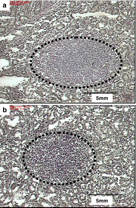
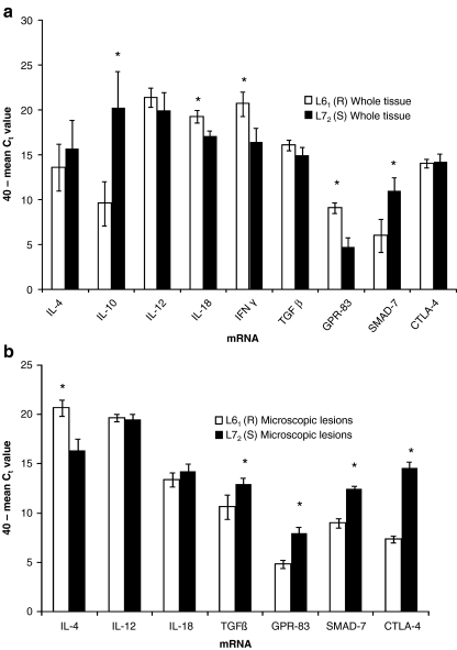
Marek's disease (MD) of chickens is a unique natural model of Hodgkin's and Non Hodgkin's lymphomas in which the neoplastically-transformed cells over-express CD30 (CD30(hi)) antigen. All chicken genotypes can be infected with MD virus and develop microscopic lymphomas. From 21 days post infection (dpi) microscopic lymphomas regress in resistant chickens but, in contrast, they progress to gross lymphomas in susceptible chickens. Here we test our hypothesis that in resistant chickens at 21 dpi the tissue microenvironment is pro T-helper (Th)-1 and compatible with cytotoxic T lymphocyte (CTL) immunity but in susceptible lines it is pro Th-2 or pro T-regulatory (T-reg) and antagonistic to CTL immunity. We used the B2, non-MHC-associated, MD resistance/susceptibility system (line [L]6(1)/line [L]7(2)) and quantified the levels of key mRNAs that can be used to define Th-1 (IL-2, IL-12, IL-18, IFNgamma), Th-2 (IL-4, IL-10) and T-reg (TGFbeta, GPR-83, CTLA-4, SMAD-7) lymphocyte phenotypes. We measured gene expression in both whole tissues (represents tissue microenvironment and tumor microenvironment) and in the lymphoma lesions (tumor microenvironment) themselves. Gene ontology-based modeling of our results shows that the dominant phenotype in whole tissue as well as in microscopic lymphoma lesions, is pro T-reg in both L6(1) and L7(2) but a minor pro Th-1 and anti Th-2 tissue microenvironment exists in L6(1) whereas there is an anti Th-1 and pro Th-2 tissue microenvironment in L7(2). The tumor microenvironment per se is pro T-reg, anti Th-1 and pro Th-2 in both L6(1) and L7(2). Together our data suggests that the neoplastic transformation is essentially the same in both L6(1) and L7(2) and that resistance/susceptibility is mediated at the level of tumor immunity in the tissues.
Figures

References
-
- Institute NC (2007) The NCI strategic plan for leading the nation to eliminate the suffering and death due to cancer. Available via: http://strategicplan.nci.nih.gov/pdf/nci_2007_strategic_plan.pdf [cited 05/29 2008]
LinkOut - more resources
Full Text Sources
Research Materials
Miscellaneous