. 2008 Dec;1(1):69-83.
doi: 10.1007/s12307-008-0007-2. Epub 2008 Mar 14.

Role of cancer microenvironment in metastasis: focus on colon cancer

Affiliations

Role of cancer microenvironment in metastasis: focus on colon cancer

Stéphanie Gout et al. Cancer Microenviron. 2008 Dec.

Abstract

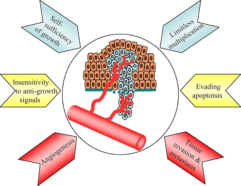
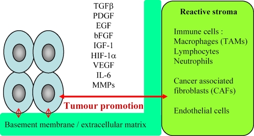
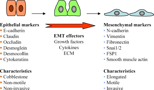
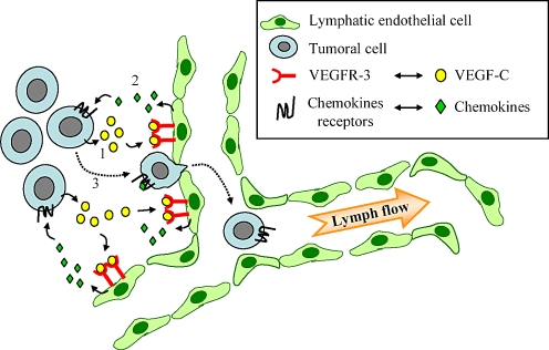
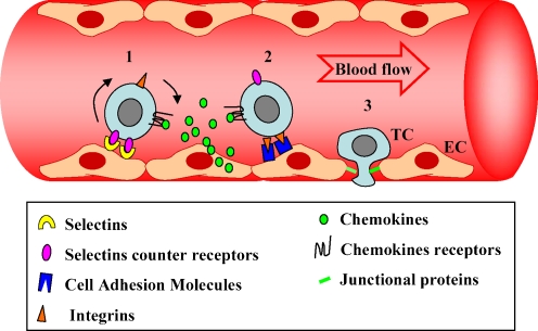
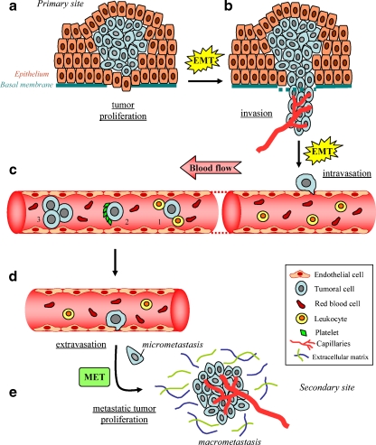
One person on three will receive a diagnostic of cancer during his life. About one third of them will die of the disease. In most cases, death will result from the formation of distal secondary sites called metastases. Several events that lead to cancer are under genetic control. In particular, cancer initiation is tightly associated with specific mutations that affect proto-oncogenes and tumour suppressor genes. These mutations lead to unrestrained growth of the primary neoplasm and a propensity to detach and to progress through the subsequent steps of metastatic dissemination. This process depends tightly on the surrounding microenvironment. In fact, several studies support the point that tumour development relies on a continuous cross-talk between cancer cells and their cellular and extracellular microenvironments. This signaling cross-talk is mediated by transmembrane receptors expressed on cancer cells and stromal cells. The aim of this manuscript is to review how the cancer microenvironment influences the journey of a metastatic cell taking liver invasion by colorectal cancer cells as a model.


Figures

Fig. 1
Fig. 1
The hallmarks of cancer cells. Cancer cells from most human cancers share six acquired traits that collectively dictate malignant growth: (a) self-sufficiency with respect to growth signals, (b) insensitivity to growth-inhibitory signals, (c) evasion of programmed cell death, (d) limitless replicative potential, (e) sustained angiogenesis, and (f) tissue invasion and metastasis. All these traits contribute to growth, detachment and invading potential of cancer cells (adapted from [1])
Fig. 2
Fig. 2
Interaction of cancer cells with the microenvironment. Beyond genetic alterations that affect cancer cells, a dynamic interaction occurs between cancer cells and the host stromal microenvironment to support cancerous growth and dissemination. The stroma is constituted mainly of cellular elements such as fibroblasts and immune cells, and non-cellular elements such as ECM. These stromal elements act in a synergistic cross-talk with the cancer cells from the primary sites to sustain cancer growth and metastasis. For example, cancer-associated macrophages and fibroblasts influence cancer initiation/promotion by secreting cytokines, growth factors and chemokines
Fig. 3
Fig. 3
The epithelial–mesenchymal transition. Epithelial-to-Mesenchymal Transition (EMT) is a morphogenetic process in which epithelial cells loose their characteristics and gain mesenchymal properties during embryogenesis and during progression of cancer. Carcinoma cells acquire a mesenchymal-like state in order to facilitate their migration and invasion. The EMT process is induced and regulated by effectors such as growth factors (TGFβ, PDGF, EGF), cytokines (Il-8) and ECM components. It is characterized by loss of epithelial markers such as E-cadherin and cytokeratins and gain of mesenchymal markers such as N-cadherin and vimentin
Fig. 4
Fig. 4
Mechanisms of VEGF-C-induced intravasation of cancer cells across lymph vessels. In several human cancers, increased expression of VEGF-C in primary tumours correlates with regional lymph node metastases. It is possible that a reciprocal cross-talk exists between tumour cells and lymphatic endothelial cells to induce tumour lymphangiogenesis and formation of lymph node metastases. Notably, VEGF-C activates lymphatic endothelial cells (1) that in turn may secrete chemotactic factors (2). This will contribute to attract cancer cells bearing appropriate chemokine receptors to the growing lymph vessels (3), and enable their adhesion and their intra-lymphatic intravasation
Fig. 5
Fig. 5
Extravasation of cancer cells is a multi-step process. The first step consists in the transient adhesion of cancer cells to the endothelium. It involves endothelial adhesion molecules such as E-selectin and P-selectin and their counter-receptors present on cancer cells. This step is associated with the rolling of the cancer cells on the endothelium (1). The second step consists in a firmer adhesion of cancer cells to endothelial cells (2). It is mediated through chemoattractants and cell adhesion molecules on the endothelium and integrins on the cancer cells. The third step is characterized by the extravasation of cancer cells through endothelial cell–cell junctions (3). EC Endothelial cells, TC tumour cell (adapted from [107])
Fig. 6
Fig. 6
The journey of a metastatic cell: from the primary site to the secondary site. Cancer cells have a “long and difficult” journey to go on before colonizing a secondary site and form metastases. This journey comprises several milestones that are summarized as follows: a Proliferation of primary tumour, b Local invasion of detached cells. c Intravasation in a capillary, and tumour cell survival in blood circulation: 1, interaction with leukocytes that may be destructive or not; 2, aggregation with platelets, which protects cancer cells against mechanical stress and leukocytes; 3, tumour cell–cell aggregation, which protects against stress and formation of intra capillary thrombosis. d Arrest and extravasation in a target organ. e Metastatic growth in the new appropriate environment and formation of a secondary neoplasm

References

    1. {'text': '', 'ref_index': 1, 'ids': [{'type': 'PubMed', 'value': '10647931', 'is_inner': True, 'url': 'https://pubmed.ncbi.nlm.nih.gov/10647931/'}]}
    2. Hanahan D, Weinberg RA (2000) The hallmarks of cancer. Cell 100:57–70 - PubMed
    1. {'text': '', 'ref_index': 1, 'ids': [{'type': 'PMC', 'value': 'PMC2001051', 'is_inner': False, 'url': 'https://pmc.ncbi.nlm.nih.gov/articles/PMC2001051/'}, {'type': 'PubMed', 'value': '17916855', 'is_inner': True, 'url': 'https://pubmed.ncbi.nlm.nih.gov/17916855/'}]}
    2. Ballinger AB, Anggiansah C (2007) Colorectal cancer. BMJ 335:715–718 - PMC - PubMed
    1. {'text': '', 'ref_index': 1, 'ids': [{'type': 'PubMed', 'value': '17243120', 'is_inner': True, 'url': 'https://pubmed.ncbi.nlm.nih.gov/17243120/'}]}
    2. Tse JC, Kalluri R (2007) Mechanisms of metastasis: epithelial-to-mesenchymal transition and contribution of tumor microenvironment. J Cell Biochem 101:816–829 - PubMed
    1. {'text': '', 'ref_index': 1, 'ids': [{'type': 'PubMed', 'value': '17226777', 'is_inner': True, 'url': 'https://pubmed.ncbi.nlm.nih.gov/17226777/'}]}
    2. Li H, Fan X, Houghton J (2007) Tumor microenvironment: the role of the tumor stroma in cancer. J Cell Biochem 101:805–815 - PubMed
    1. {'text': '', 'ref_index': 1, 'ids': [{'type': 'PubMed', 'value': '12663862', 'is_inner': True, 'url': 'https://pubmed.ncbi.nlm.nih.gov/12663862/'}]}
    2. Mareel M, Leroy A (2003) Clinical, cellular, and molecular aspects of cancer invasion. Physiol Rev 83:337–376 - PubMed

LinkOut - more resources