The prometastatic microenvironment of the liver
- PMID: 19308690
- PMCID: PMC2654354
- DOI: 10.1007/s12307-008-0011-6
The prometastatic microenvironment of the liver
Abstract
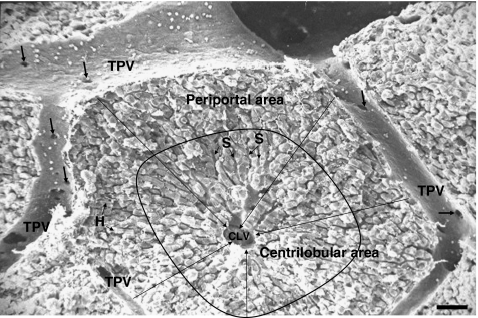
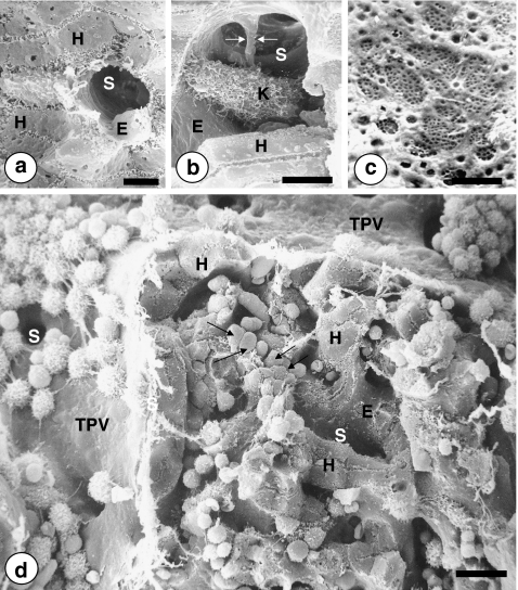
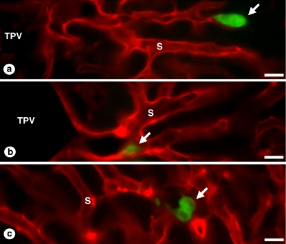
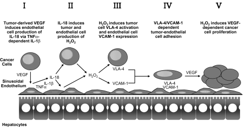
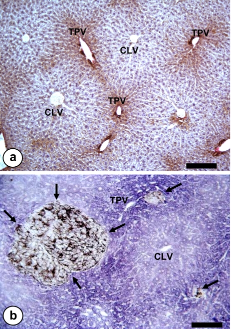
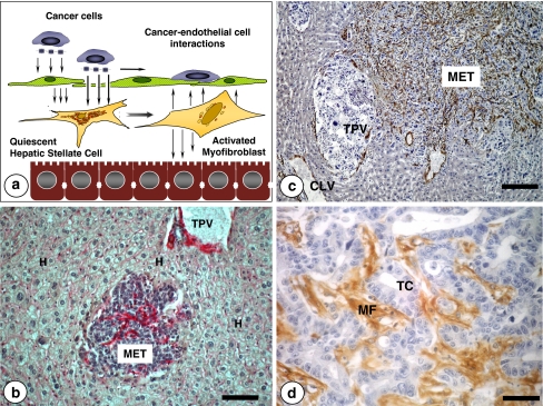
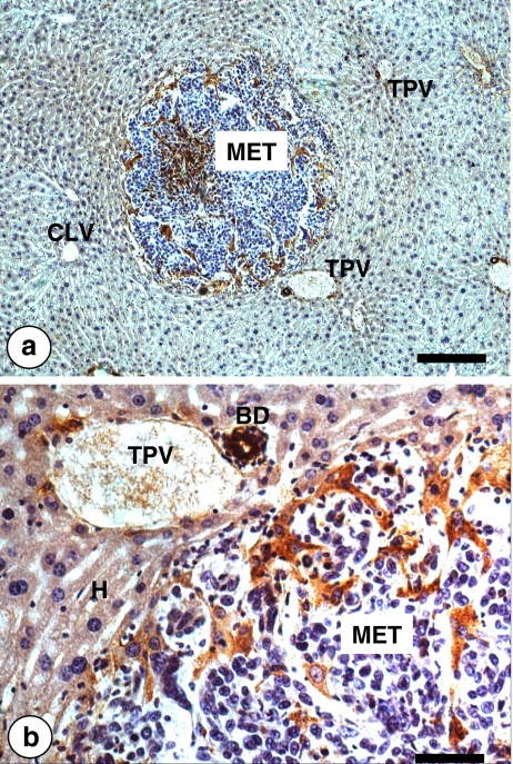
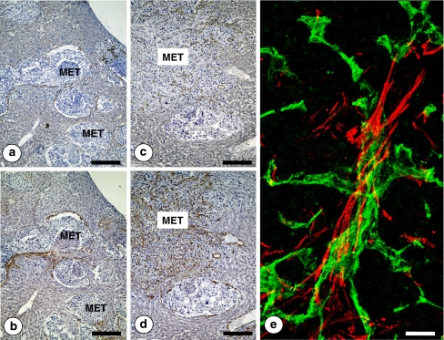
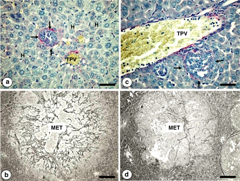
The liver is a major metastasis-susceptible site and majority of patients with hepatic metastasis die from the disease in the absence of efficient treatments. The intrahepatic circulation and microvascular arrest of cancer cells trigger a local inflammatory reaction leading to cancer cell apoptosis and cytotoxicity via oxidative stress mediators (mainly nitric oxide and hydrogen peroxide) and hepatic natural killer cells. However, certain cancer cells that resist or even deactivate these anti-tumoral defense mechanisms still can adhere to endothelial cells of the hepatic microvasculature through proinflammatory cytokine-mediated mechanisms. During their temporary residence, some of these cancer cells ignore growth-inhibitory factors while respond to proliferation-stimulating factors released from tumor-activated hepatocytes and sinusoidal cells. This leads to avascular micrometastasis generation in periportal areas of hepatic lobules. Hepatocytes and myofibroblasts derived from portal tracts and activated hepatic stellate cells are next recruited into some of these avascular micrometastases. These create a private microenvironment that supports their development through the specific release of both proangiogenic factors and cancer cell invasion- and proliferation-stimulating factors. Moreover, both soluble factors from tumor-activated hepatocytes and myofibroblasts also contribute to the regulation of metastatic cancer cell genes. Therefore, the liver offers a prometastatic microenvironment to circulating cancer cells that supports metastasis development. The ability to resist anti-tumor hepatic defense and to take advantage of hepatic cell-derived factors are key phenotypic properties of liver-metastasizing cancer cells. Knowledge on hepatic metastasis regulation by microenvironment opens multiple opportunities for metastasis inhibition at both subclinical and advanced stages. In addition, together with metastasis-related gene profiles revealing the existence of liver metastasis potential in primary tumors, new biomarkers on the prometastatic microenvironment of the liver may be helpful for the individual assessment of hepatic metastasis risk in cancer patients.
Figures


Similar articles
-
Cooperation of liver cells in health and disease.Adv Anat Embryol Cell Biol. 2001;161:III-XIII, 1-151. doi: 10.1007/978-3-642-56553-3. Adv Anat Embryol Cell Biol. 2001. PMID: 11729749 Review.
-
The liver prometastatic reaction of cancer patients: implications for microenvironment-dependent colon cancer gene regulation.Cancer Microenviron. 2011 Aug;4(2):163-80. doi: 10.1007/s12307-011-0084-5. Epub 2011 Aug 26. Cancer Microenviron. 2011. PMID: 21870094 Free PMC article.
-
Proangiogenic role of tumor-activated hepatic stellate cells in experimental melanoma metastasis.Hepatology. 2003 Mar;37(3):674-85. doi: 10.1053/jhep.2003.50068. Hepatology. 2003. PMID: 12601365
-
Hydrogen peroxide mediates vascular cell adhesion molecule-1 expression from interleukin-18-activated hepatic sinusoidal endothelium: implications for circulating cancer cell arrest in the murine liver.Hepatology. 2001 Aug;34(2):298-310. doi: 10.1053/jhep.2001.26629. Hepatology. 2001. PMID: 11481615
-
Liver prometastatic reaction: Stimulating factors and responsive cancer phenotypes.Semin Cancer Biol. 2021 Jun;71:122-133. doi: 10.1016/j.semcancer.2020.08.001. Epub 2020 Aug 15. Semin Cancer Biol. 2021. PMID: 32805395 Review.
Cited by
-
Dual Role of Fibroblasts Educated by Tumour in Cancer Behavior and Therapeutic Perspectives.Int J Mol Sci. 2022 Dec 8;23(24):15576. doi: 10.3390/ijms232415576. Int J Mol Sci. 2022. PMID: 36555218 Free PMC article. Review.
-
CDK8 Selectively Promotes the Growth of Colon Cancer Metastases in the Liver by Regulating Gene Expression of TIMP3 and Matrix Metalloproteinases.Cancer Res. 2018 Dec 1;78(23):6594-6606. doi: 10.1158/0008-5472.CAN-18-1583. Epub 2018 Sep 5. Cancer Res. 2018. PMID: 30185549 Free PMC article.
-
The Tumor Microenvironment in Liver Metastases from Colorectal Carcinoma in the Context of the Histologic Growth Patterns.Int J Mol Sci. 2021 Feb 3;22(4):1544. doi: 10.3390/ijms22041544. Int J Mol Sci. 2021. PMID: 33546502 Free PMC article. Review.
-
Invasion and metastasis in cancer: molecular insights and therapeutic targets.Signal Transduct Target Ther. 2025 Feb 21;10(1):57. doi: 10.1038/s41392-025-02148-4. Signal Transduct Target Ther. 2025. PMID: 39979279 Free PMC article. Review.
-
The type I insulin-like growth factor regulates the liver stromal response to metastatic colon carcinoma cells.Oncotarget. 2016 Oct 12;8(32):52281-52293. doi: 10.18632/oncotarget.12595. eCollection 2017 Aug 8. Oncotarget. 2016. PMID: 28881729 Free PMC article.
References
-
- None
- Pickren JW, Tsukada Y, Lane WW (1982) Liver metastasis. In: Weiss L, Gilbert HA (eds) Analysis of autopsy data. GK Hall and Company, Boston, Mass, pp 2–18
-
- None
- Gilbert HA, Kagan AR, Hintz BL et al (1982) Patterns of metastases. In: Weiss L, Gilbert HA (eds) Liver metastases. GK Hall Medical Publishers, Boston, MA, pp 19–39
-
- {'text': '', 'ref_index': 1, 'ids': [{'type': 'DOI', 'value': '10.1038/nrc1098', 'is_inner': False, 'url': 'https://doi.org/10.1038/nrc1098'}, {'type': 'PubMed', 'value': '12778135', 'is_inner': True, 'url': 'https://pubmed.ncbi.nlm.nih.gov/12778135/'}]}
- Fidler IJ (2003) The pathogenesis of cancer metastasis: the ‘seed and soil’ hypothesis revisited. Nat Rev Cancer 3:453–458 - PubMed
-
- None
- Ewing J (1928) Neoplastic diseases, 6th edn. WB Saunders, Philadelphia
-
- {'text': '', 'ref_index': 1, 'ids': [{'type': 'DOI', 'value': '10.1016/S0140-6736(00)49915-0', 'is_inner': False, 'url': 'https://doi.org/10.1016/s0140-6736(00)49915-0'}, {'type': 'PubMed', 'value': '2673568', 'is_inner': True, 'url': 'https://pubmed.ncbi.nlm.nih.gov/2673568/'}]}
- Paget S (1889) The distribution of secondary growths in cancer of the breast. Lancet 1:571–573 - PubMed
LinkOut - more resources
Full Text Sources