

Sialyl Lewis X expression and lymphatic microvessel density in primary tumors of node-negative colorectal cancer patients predict disease recurrence
- PMID: 19308692
- PMCID: PMC2654349
- DOI: 10.1007/s12307-008-0014-3
Sialyl Lewis X expression and lymphatic microvessel density in primary tumors of node-negative colorectal cancer patients predict disease recurrence
Abstract
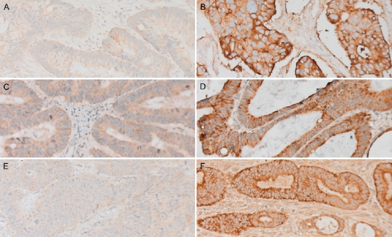
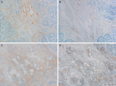
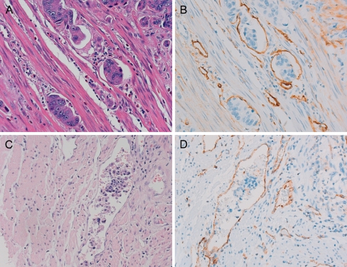
Up to 30% of curatively resected colorectal cancer patients with tumor-negative lymph nodes, show disease recurrence. We assessed whether these high-risk patients can be identified by examining primary tumors for the following blood and lymphatic vasculature markers: A) sialyl Lewis X (sLeX), vascular endothelial growth factor (VEGF)-C and VEGF-D expression; B) blood and lymphatic microvessel density (BMVD/LMVD); and C) the presence of blood and lymphatic vessel invasion. Thirty-six cases (disease recurrence within 5 years) and 72 controls (no disease recurrence for at least 5 years) were selected in a case-control design. Tumor sections were stained by antibodies CSLEX1 (sLeX), anti-VEGF-C, anti-VEGF-D, anti-CD31 (BMVD) or D2-40 (LMVD) to determine the parameters as mentioned above. A multivariate analysis showed sLeX expression and high LMVD (odds ratio 5.1, 95% confidence interval 1.3-20.0 and odds ratio 3.1, 95% confidence interval 1.0-10.0, respectively) to be independent factors predicting disease recurrence. Expression of sLeX correlated with liver metastases (P = 0.015). A high LMVD was related to regional intra-abdominal or intrapelvic metastases in lymph nodes and distant metastases other than in the liver and lungs such as peritoneum, bones, brain and adrenal glands (P = 0.004). A high BMVD in the invasive front correlated with lung metastases (P = 0.018). We show that high-risk node-negative colorectal cancer patients can be identified by primary tumor assessment for sLeX expression and LMVD. Our results are consistent with the notion that both lymphatic and hematogenous metastasis play a role in colorectal cancer.
Figures
Similar articles
-
[Clinical significance of detection on lymphatic microvessel, lymphatic microvessel density and vascular endothelial growth factor-C in patients with colorectal carcinoma].Zhonghua Wei Chang Wai Ke Za Zhi. 2006 Nov;9(6):477-82. Zhonghua Wei Chang Wai Ke Za Zhi. 2006. PMID: 17143789 Chinese.
-
Lymphatic microvessel density as a predictive marker for the recurrence time of pterygium: a three-year follow-up study.Mol Vis. 2013;19:166-73. Epub 2013 Jan 28. Mol Vis. 2013. PMID: 23378730 Free PMC article.
-
Prognostic significance of VEGF-C expression in correlation with COX-2, lymphatic microvessel density, and clinicopathologic characteristics in human non-small cell lung cancer.Acta Biochim Biophys Sin (Shanghai). 2009 Mar;41(3):217-22. doi: 10.1093/abbs/gmp004. Acta Biochim Biophys Sin (Shanghai). 2009. PMID: 19280060
-
A meta-analysis of the relationship between lymphatic microvessel density and clinicopathological parameters in breast cancer.Bull Cancer. 2013 Mar;100(3):1-10. doi: 10.1684/bdc.2013.1719. Bull Cancer. 2013. PMID: 23501839 Review.
-
Clinicopathological and prognostic significance of blood microvessel density in endometrial cancer: a meta-analysis and subgroup analysis.Arch Gynecol Obstet. 2018 Mar;297(3):731-740. doi: 10.1007/s00404-018-4648-1. Epub 2018 Jan 11. Arch Gynecol Obstet. 2018. PMID: 29327157 Review.
Cited by
-
Multivalent conjugation of antibody to dendrimers for the enhanced capture and regulation on colon cancer cells.Sci Rep. 2015 Mar 30;5:9445. doi: 10.1038/srep09445. Sci Rep. 2015. PMID: 25819426 Free PMC article.
-
Associated expression of α2,3sialylated type 2 chain structures with lymph node metastasis in distal colorectal cancer.Surg Today. 2013 Feb;43(2):155-62. doi: 10.1007/s00595-012-0141-9. Epub 2012 Mar 8. Surg Today. 2013. PMID: 22398718
-
The Expression of CD10 and CD15 Is Progressively Increased during Colorectal Cancer Development.Korean J Pathol. 2013 Aug;47(4):340-7. doi: 10.4132/KoreanJPathol.2013.47.4.340. Epub 2013 Aug 26. Korean J Pathol. 2013. PMID: 24009629 Free PMC article.
-
Lymphangiogenesis and colorectal cancer.Saudi Med J. 2017 Mar;38(3):237-244. doi: 10.15537/smj.2017.3.16245. Saudi Med J. 2017. PMID: 28251217 Free PMC article. Review.
-
ING4 is negatively correlated with microvessel density in colon cancer.Tumour Biol. 2012 Dec;33(6):2357-64. doi: 10.1007/s13277-012-0498-9. Epub 2012 Sep 28. Tumour Biol. 2012. PMID: 23055189
References
-
- {'text': '', 'ref_index': 1, 'ids': [{'type': 'PubMed', 'value': '2300087', 'is_inner': True, 'url': 'https://pubmed.ncbi.nlm.nih.gov/2300087/'}]}
- Moertel CG, Fleming TR, Macdonald JS et al (1990) Levamisole and fluorouracil for adjuvant therapy of resected colon carcinoma. N Engl J Med 322:352–358 - PubMed
-
- {'text': '', 'ref_index': 1, 'ids': [{'type': 'PubMed', 'value': '10334519', 'is_inner': True, 'url': 'https://pubmed.ncbi.nlm.nih.gov/10334519/'}]}
- Impact B2 Investigators (1999) Efficacy of adjuvant fluorouracil and folinic acid in B2 colon cancer. International Multicentre Pooled Analysis of B2 Colon Cancer Trials (IMPACT B2) Investigators. J Clin Oncol 17:1356–1363 - PubMed
-
- {'text': '', 'ref_index': 1, 'ids': [{'type': 'DOI', 'value': '10.1007/s00384-006-0098-5', 'is_inner': False, 'url': 'https://doi.org/10.1007/s00384-006-0098-5'}, {'type': 'PubMed', 'value': '16528541', 'is_inner': True, 'url': 'https://pubmed.ncbi.nlm.nih.gov/16528541/'}]}
- Madbouly KM, Senagore AJ, Mukerjee A et al (2007) Does immunostaining effectively upstage colorectal cancer by identifying micrometastatic nodal disease? Int J Colorectal Dis 22:39–48 - PubMed
-
- {'text': '', 'ref_index': 1, 'ids': [{'type': 'DOI', 'value': '10.1007/s00268-001-0236-8', 'is_inner': False, 'url': 'https://doi.org/10.1007/s00268-001-0236-8'}, {'type': 'PubMed', 'value': '11865379', 'is_inner': True, 'url': 'https://pubmed.ncbi.nlm.nih.gov/11865379/'}]}
- Cianchi F, Palomba A, Boddi V et al (2002) Lymph node recovery from colorectal tumor specimens: recommendation for a minimum number of lymph nodes to be examined. World J Surg 26:384–389 - PubMed
-
- {'text': '', 'ref_index': 1, 'ids': [{'type': 'DOI', 'value': '10.1016/S0140-6736(07)61058-7', 'is_inner': False, 'url': 'https://doi.org/10.1016/s0140-6736(07)61058-7'}, {'type': 'PubMed', 'value': '18083404', 'is_inner': True, 'url': 'https://pubmed.ncbi.nlm.nih.gov/18083404/'}]}
- Quasar Collaborative Group, Gray R, Barnwell J et al (2007) Adjuvant chemotherapy versus observation in patients with colorectal cancer: a randomised study. Lancet 370:2020–2029 - PubMed
LinkOut - more resources
Full Text Sources