. 2009 Jun;30(6):1791-800.
doi: 10.1002/hbm.20763.

Prestimulus alpha and mu activity predicts failure to inhibit motor responses

Affiliations

Prestimulus alpha and mu activity predicts failure to inhibit motor responses

Ali Mazaheri et al. Hum Brain Mapp. 2009 Jun.

Abstract

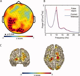
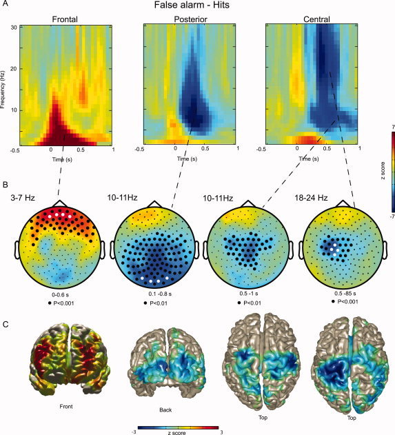
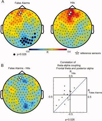
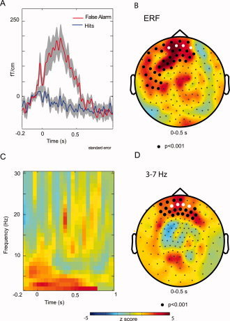
Do certain brain states predispose humans to commit errors in monotonous tasks? We used MEG to investigate how oscillatory brain activity indexes the brain state in subjects performing a Go-noGo task. Elevated occipital alpha and sensorimotor mu activity just prior to the presentation of the stimuli predicted an upcoming error. An error resulted in increased frontal theta activity and decreased posterior alpha activity. This theta increase and alpha decrease correlated on a trial-by-trial basis reflecting post-error functional connectivity between the frontal and occipital regions. By examining the state of the brain before a stimulus, we were able to show that it is possible to predict lapses of attention before they actually occur. This supports the case that the state of the brain is important for how incoming stimuli are processed and for how subjects respond.


Figures

Figure 1
Figure 1
The power calculated in prestimulus interval (time −1 to 0 s) for False Alarms compared to Correct Withholds. (A) Topography of the 10–11 Hz power of the difference between False Alarms and Correct Withholds averaged over subjects (planar gradient). The cluster of sensors showing significantly stronger alpha power for False Alarms than Correct Withholds is marked with dots (P < 0.008; cluster randomization routine). (B) Grand average of the spectra calculated (−1 to 0 s; red line, False Alarms; blue line, Correct Withholds). The spectra were averaged over the cluster of sensors that showed a significant difference. (C) Using a beamforming approach we identified the regions accounting for the difference in alpha power between False Alarms and Correct Rejections to occipital and sensorimotor cortex.
Figure 2
Figure 2
The power of oscillatory activity characterized in the postresponse interval for False Alarms compared to Hits. (A) Grand average of the difference TFRs (False Alarms–Hits) of representative frontal, posterior and central sensors (sensors marked in white). The button press occurred at t = 0 s. No baseline correction was applied. (B) Grand average of the topography of theta (3–7 Hz), alpha (10–11 Hz), mu (10–11 Hz), and beta (18–24 Hz) activity. The dots denote clusters representing significant differences (cluster randomization routine). (C) A beamforming technique was applied to localize the regions responsible for producing the difference in the power shown in (B). The theta increase was localized to frontal regions. Decrease in alpha and mu activity was localized to occipital cortex and extended sensorimotor areas. The beta decrease was localized to primary sensorimotor areas.
Figure 3
Figure 3
Correlations between the frontal theta increase and the posterior alpha decrease. Two frontal sensors (marked with stars) were used a reference for the correlation analysis. The correlations between theta power in the reference sensors and the alpha power in all the other sensors were calculated on a trial‐by‐trial basis (0–500 ms after response). (A) Left panel: grand average of the theta‐alpha correlation for False Alarms. Note the strong anticorrelation in an isolated region over posterior regions (P < 0.028, one sample t‐test, cluster randomization routine). Right panel: grand average of the theta‐alpha correlations for Hits. There were no significant correlations. (B) The difference in theta‐alpha power correlations between False Alarms and Hits. Right panel, the theta‐alpha power correlation of the 14 subjects for False Alarms (x‐axis) and Hits (y‐axis). They were calculated for the two frontal sensors (marked by stars) and the posterior significant sensors (A, right panel). The correlations over the right posterior sensors were significantly more negative for False Alarm than Hits (P < 0.026, pair‐wise t‐test).
Figure 4
Figure 4
The time‐locked signals in the postresponse interval. (A) The ERFs with respect to False Alarms (red) and Hits (blue) for representative frontal sensors (marked in white). Button press was a t = 0 s. (B) The topography of the ERF difference between False Alarms and Hits (0–0.5 s). Even though the difference was dominated by frontal sensors, the cluster of sensors representing the significant difference (P < 0.001; cluster randomization routine) was large. (C) The difference in TFRs of the ERFs for False Alarms and Hits. (D) The topography of the difference in TFRs of the ERFs. Note the frontal distribution of the significant cluster (P < 0.001; cluster randomization routine).

References

    1. Ahonen AI,Hamalainen MS,Ilmoniemi RJ,Kajola MJ,Knuutila JE,Simola JT,Vilkman VA ( 1993): Sampling theory for neuromagnetic detector arrays. IEEE Trans Biomed Eng 40: 859–869. - PubMed
    1. Bastiaansen MC,Knosche TR ( 2000): Tangential derivative mapping of axial MEG applied to event‐related desynchronization research. Clin Neurophysiol 111: 1300–1305. - PubMed
    1. Bauer M,Oostenveld R,Peeters M,Fries P ( 2006): Tactile spatial attention enhances gamma‐band activity in somatosensory cortex and reduces low‐frequency activity in parieto‐occipital areas. J Neurosci 26: 490–501. - PMC - PubMed
    1. Bokura H,Yamaguchi S,Kobayashi S ( 2001): Electrophysiological correlates for response inhibition in a Go/NoGo task. Clin Neurophysiol 112: 2224–2232. - PubMed
    1. Bray S,O'Doherty J ( 2007): Neural coding of reward‐prediction error signals during classical conditioning with attractive faces. J Neurophysiol 97: 3036–3045. - PubMed

Publication types