. 2009 Apr 14;48(14):3100-8.
doi: 10.1021/bi801750u.

Characterization of peptidoglycan in fem-deletion mutants of methicillin-resistant Staphylococcus aureus by solid-state NMR

Affiliations

Characterization of peptidoglycan in fem-deletion mutants of methicillin-resistant Staphylococcus aureus by solid-state NMR

Shasad Sharif et al. Biochemistry. .

Abstract

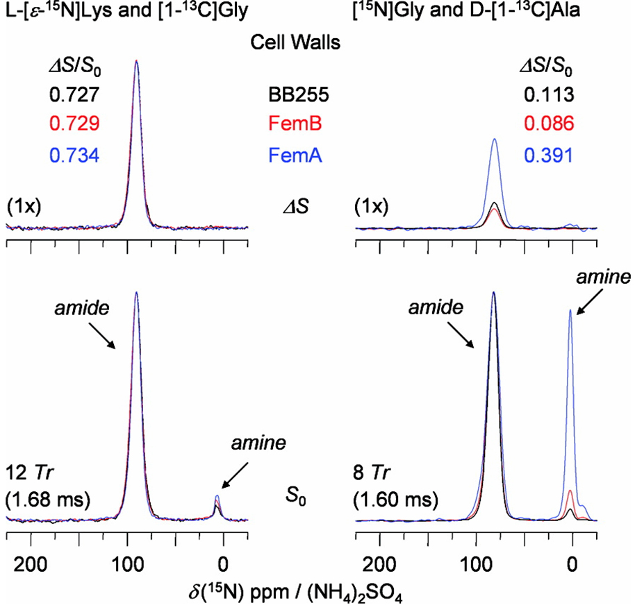
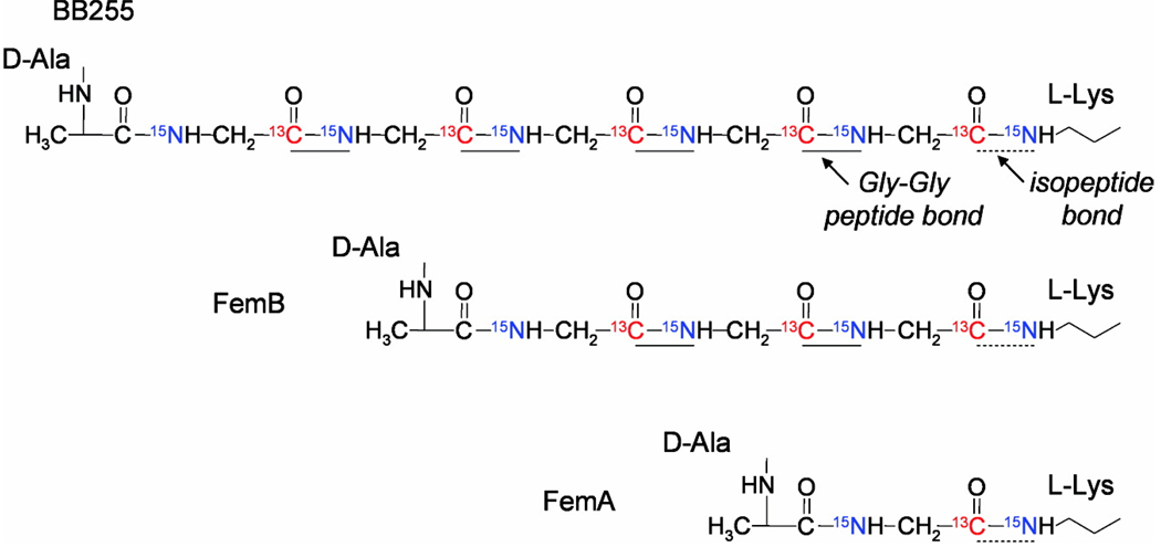
Compositional analysis of the peptidoglycan (PG) of a wild-type methicillin-resistant Staphylococcus aureus and its fem-deletion mutants has been performed on whole cells and cell walls using stable-isotope labeling and rotational-echo double-resonance NMR. The labels included [1-(13)C,(15)N]glycine and l-[epsilon-(15)N]lysine (for a direct measure of the number of glycyl residues in the bridging segment), [1-(13)C]glycine and l-[epsilon-(15)N]lysine (concentration of bridge links), and d-[1-(13)C]alanine and [(15)N]glycine (concentrations of cross-links and wall teichoic acids). The bridging segment length changed from 5.0 glycyl residues (wild-type strain) to 2.5 +/- 0.1 (FemB) with modest changes in cross-link and bridge-link concentrations. This accurate in situ measurement for the FemB mutant indicates a heterogeneous PG structure with 25% monoglycyl and 75% triglycyl bridges. When the bridging segment was reduced to a single glycyl residue 1.0 +/- 0.1 (FemA), the level of cross-linking decreased by more than 20%, resulting in a high concentration of open N-terminal glycyl segments.


Figures

Figure 1
Figure 1
Transmission electron micrographs of whole-cells of S. aureus grown in defined medium (SASM) and harvested at O.D. 0.2 at 660 nm. (Left) wild-type BB255. (Middle) FemB (UT34-2). (Right) FemA (UK 17). The arrows indicate morphological irregularities for the mutant strains. Average cell size for the mutants did not exceed that of the wild-type strain.
Figure 2
Figure 2
13C{15N} REDOR spectra after 8-Tr dipolar evolution (1.60 ms) obtained at 75 MHz with 5-kHz MAS of whole cells of wild-type S. aureus (BB255, black), and FemB (UT 34-2, red) and FemA (UK 17, blue) mutants labeled by [1-13C, 15N]glycine and L-[ε-15N]lysine. The full-echo spectra (S0) are at the bottom of the figure, and the REDOR differences (ΔS = S0S, where S is the dephased echo) are shown at the top. Natural-abundance carbon peaks appear between 20–70 ppm in the S0 spectrum (bottom inset). The 171-ppm peak intensity in the ΔS spectrum (top inset) of wild-type S. aureus BB255 was normalized to a five glycine-equivalent unit (GEU). The 13C shoulder for FemA (top inset) is assigned to glycyl amines of open bridging segments with terminal NH2 groups. The spectra were scaled according to sample weight and total number of scans; 13C chemical shifts are with respect to external tetramethylsilane TMS.
Figure 3
Figure 3
15N{13C} REDOR spectra of cell-wall isolates of wild-type S. aureus (BB255, black) and its FemB (UT 34-2, red) and FemA (UK 17, blue) mutants labeled by (left) L-[ε-15N]lysine and [1-13C]glycine, and (right) [15N]glycine and D-[1-13C]alanine. The full-echo spectra (S0) are at the bottom of the figure, and the REDOR differences (ΔS = S0S, where S is the dephased echo) are shown at the top. The spectra on the left were obtained at 50 MHz with 7143-Hz MAS. After an evolution time of 1.68 ms, the amide peak at ~92 ppm is due to a lysyl bridge-link, and the amine peak at ~9 ppm is from an [ε-15N]lysyl residue that is not bridge-linked. The spectra on the right were obtained at 30 MHz with 5-kHz MAS. After an evolution time of 1.60 ms, the amide peak at ~90 ppm is due to a glycyl-D-alanyl cross-link, and the amine peak at ~8 ppm is from a terminal glycyl bridging segment that is not cross-linked. 15N chemical shifts are with respect to external ammonium sulfate (NH4)2SO4.
Scheme 1
Scheme 1
(Left) Schematic representation of an idealized cell-wall peptdioglycan of Staphylococcus aureus and its fem-deletion mutants. A four unit peptide stem (triangles) having the sequence, L-Ala-D-iGln-L-Lys-D-Ala, is attached to every second sugar of the glycan backbone (open circles). Cross-linking between glycans occurs through glycyl side chains (dark circles) connecting the carbonyl-carbon of D-Ala of the fourth position of one stem to the ε-nitrogen (in an isopeptide bond) of L-Lys of the third position of another. The glycine content in the side chain varies from pentaglycyl (S. aureus), triglycyl (FemB), monoglycyl (FemA), to an accumulation of unsubstituted precursors with no bridges (FemX). (Right) Chemical structure of the peptidoglycan stems in the peptidoglycan of Staphylococcus aureus and its fem-deletion mutants.
Scheme 2
Scheme 2
Distribution of labels in the cell-wall peptidoglycan of a wild-type Staphylococcus aureus, and its FemB and FemA mutants, grown in defined medium containing [1-13C,15N]glycine and L-[ε-15N]lysine.

Similar articles

Cited by

References

    1. Hiramatsu K, Cui L, Kuroda M, Ito T. The emergence and evolution of methicillin-resistant Staphylococcus aureus. Trends Microbiol. 2001;9:486–493. - PubMed
    1. Labischinski H. Consequences of the interaction of β-lactam antibiotics with penicillin-binding proteins from sensitive and resistant Staphylococcus aureus strains. Med. Microbiol. Immunol. 1992;181:241–265. - PubMed
    1. Ubukata K, Nonoguchi R, Matsuhashi M, Konno M. Expression and inducibility in Staphylococcus aureus of the mecA gene, which encodes a methicillin-resistant S. aureus-specific penicillin-binding protein. J. Bacteriol. 1989;171:2882–2885. - PMC - PubMed
    1. Rohrer S, Berger-Bächi B. FemABX peptidyl transferases: a link between branched-chain cell wall peptide formation and β-lactam resistance in Gram-positive cocci. Antimicrob. Agents Ch. 2003;47:837–846. - PMC - PubMed
    1. Berger-Bächi B, Tschierske M. Role of Fem factors in methicillin resistance. Drug Resist. Updates. 1998;1:325–335. - PubMed

Publication types

MeSH terms