. 2009 Oct;12(9):1209-21.
doi: 10.1017/S1461145709000200. Epub 2009 Mar 24.

Chronic vagus nerve stimulation induces neuronal plasticity in the rat hippocampus

Affiliations

Chronic vagus nerve stimulation induces neuronal plasticity in the rat hippocampus

Francesca Biggio et al. Int J Neuropsychopharmacol. 2009 Oct.

Abstract

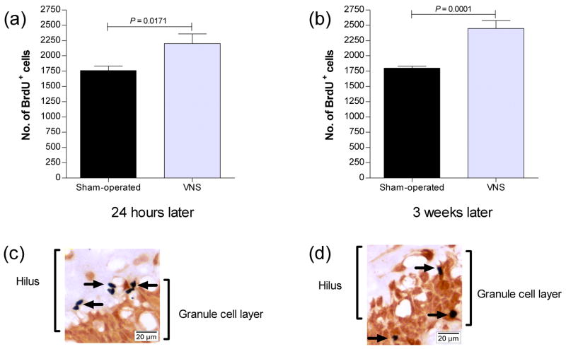
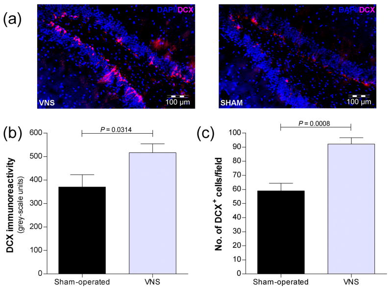
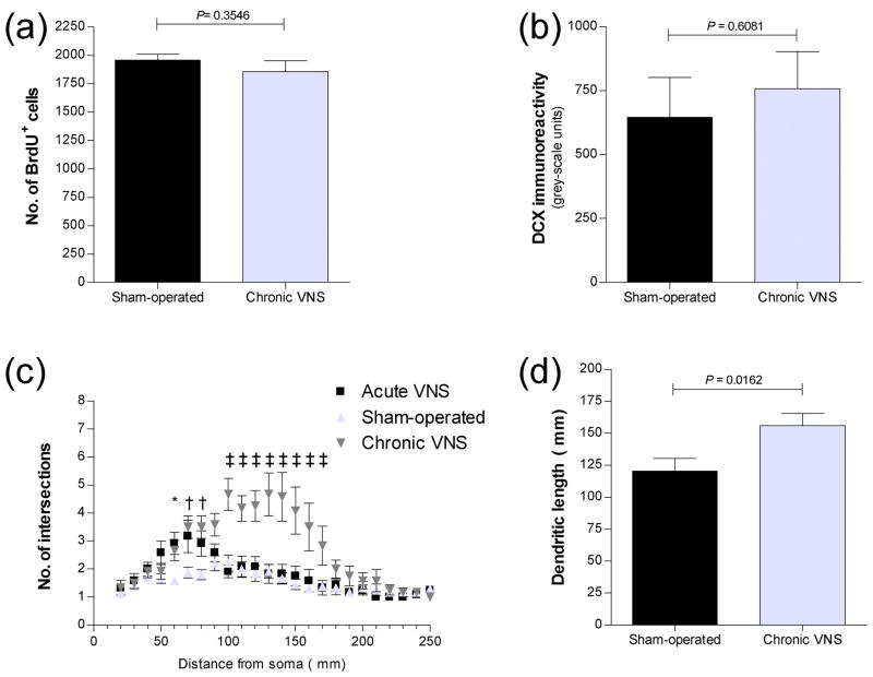
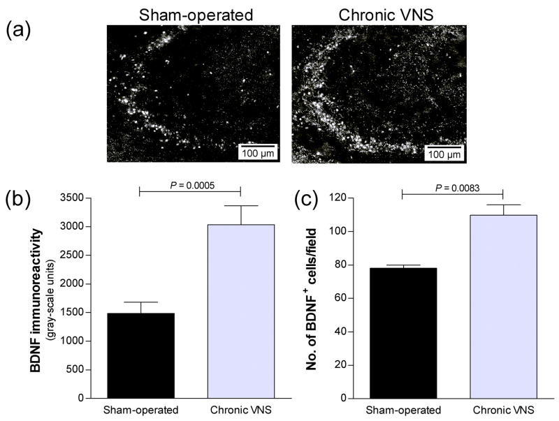
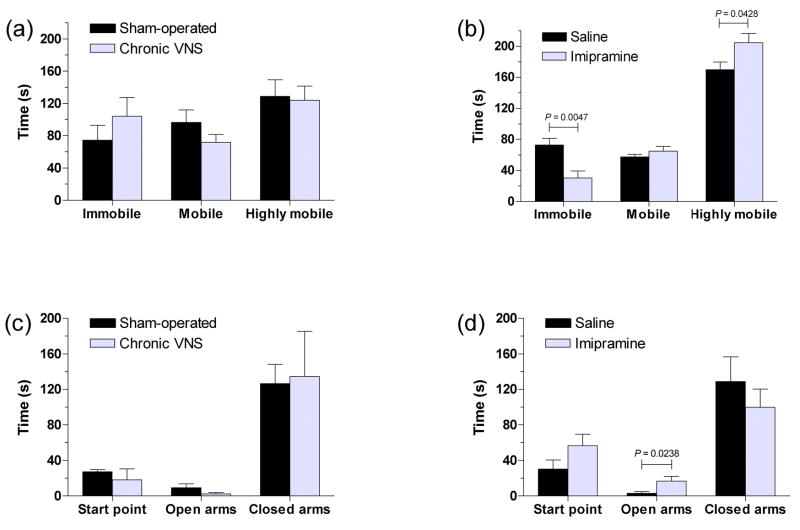
Vagus nerve stimulation (VNS) is used to treat pharmacotherapy-resistant epilepsy and depression. However, the mechanisms underlying the therapeutic efficacy of VNS remain unclear. We examined the effects of VNS on hippocampal neuronal plasticity and behaviour in rats. Cell proliferation in the hippocampus of rats subjected to acute (3 h) or chronic (1 month) VNS was examined by injection of bromodeoxyuridine (BrdU) and immunohistochemistry. Expression of doublecortin (DCX) and brain-derived neurotrophic factor (BDNF) was evaluated by immunofluorescence staining. The dendritic morphology of DCX+ neurons was measured by Sholl analysis. Our results show that acute VNS induced an increase in the number of BrdU+ cells in the dentate gyrus that was apparent 24 h and 3 wk after treatment. It also induced long-lasting increases in the amount of DCX immunoreactivity and in the number of DCX+ neurons. Neither the number of BrdU+ cells nor the amount of DCX immunoreactivity was increased 3 wk after the cessation of chronic VNS. Chronic VNS induced long-lasting increases in the amount of BDNF immunoreactivity and the number of BDNF+ cells as well as in the dendritic complexity of DCX+ neurons in the hippocampus. In contrast to chronic imipramine treatment, chronic VNS had no effect on the behaviour of rats in the forced swim or elevated plus-maze tests. Both chronic and acute VNS induced persistent changes in hippocampal neurons that may play a key role in the therapeutic efficacy of VNS. However, these changes were not associated with evident behavioural alterations characteristic of an antidepressant or anxiolytic action.


Conflict of interest statement

Statement of Interest

None

Figures

Figure 1
Figure 1
Quantitation of newly generated cells in the dentate gyrus of the rat hippocampal formation after acute VNS. (a and b) The number of BrdU+ cells in the subgranular zone and granule cell layer of the dorsal dentate gyrus was determined 24 h (a) or 3 weeks (b) after administration of VNS for 3 h by multiplying by 6 the number of positive cells counted in 24 sections with a thickness of 16 μm and spaced 96 μm apart per rat. Data are means ± SEM of values from six rats per group. P values for comparison between VNS-treated and sham-operated animals were determined by ANOVA followed by Scheffe’s test. (c and d) Representative bright-field images of immunohistochemical staining for both BrdU and NeuN in sections of the dorsal dentate gyrus obtained from rats 24 h (c) or 3 weeks (d) after acute VNS stimulation. Newly generated cells, the nuclei of which were stained dark blue for BrdU immunoreactivity (arrows), were detected in the subgranular zone in (c) and in the inner granule cell layer (stained red for NeuN immunoreactivity) in (d). Scale bars, 20 μm.
Figure 2
Figure 2
Increase in the number of DCX+ neurons in the dentate gyrus of the rat hippocampal formation induced by acute VNS. (a) Immunofluorescence staining for DCX (red) and nuclear staining with DAPI (blue) in the two blades of the granule cell layer of the dorsal dentate gyrus obtained from rats 3 weeks after acute VNS or from sham-operated controls. Scale bar, 100 μm. (b and c) Quantitation of DCX immunoreactivity (b) and the number of DCX+ neurons per field (c) in the dorsal dentate gyrus of rats treated as in (a) was performed as described in Methods and Materials. Data are means ± SEM of values from six rats per group. P values for the indicated comparisons were determined by ANOVA followed by Scheffe’s test.
Figure 3
Figure 3
Quantitation of newly generated cells (a) and effects of chronic VNS on the dendritic morphology of DCX+ neurons (b–f) in the dentate gyrus of the rat hippocampal formation after chronic VNS. (a) The number of BrdU+ cells in the subgranular zone and granule cell layer of the dorsal dentate gyrus was determined (as in Figure 1) 3 weeks after administration of VNS for 1 month. Data are means ± SEM of values from six rats per group. The P value for comparison between VNS-treated and sham-operated animals was determined by ANOVA followed by Scheffe’s test. (b) Quantitation of DCX immunoreactivity in the dorsal dentate gyrus 3 weeks after chronic VNS for 1 month. Data are means ± SEM of values from six rats per group. The P value for comparison with sham-operated animals was determined by ANOVA followed by Scheffe’s test. (c) Sholl analysis of apical dendrites of DCX+ neurons in the dorsal dentate gyrus of rats 3 weeks after acute or chronic VNS. The numbers of dendrites that cross the indicated radial distances (0 to 250 μm) from the soma are shown. Data are means ± SEM of values from six rats per group. *P < 0.05, †P < 0.01, ‡P < 0.001 versus corresponding sham-operated controls (Newman-Keuls test). (d) Dendritic length for DCX+ neurons in the dorsal dentate gyrus of rats 3 weeks after chronic VNS. Data are means ± SEM of values from six rats per group. The P value for comparison with sham-operated controls was determined by ANOVA followed by Scheffe’s test. (e–f) Representative immunofluorescence images of neurons positive for DCX or NeuN in the dentate gyrus of the rat hippocampal formation after chronic VNS. Sections of the dorsal dentate gyrus obtained from rats 3 weeks after chronic VNS (f) or from sham-operated controls (e) were stained with antibodies to DCX (red) or to NeuN (green). The boxed regions in the top panels are shown at higher magnification in the bottom panels. Note the increase in dendritic complexity and length for DCX+ neurons in rats subjected to VNS compared with those in control animals. The dendrites project deeply into the hippocampal molecular layer through the granule cell layer stained with the neuronal marker NeuN. Arrows in the merged images indicate that most DCX+ neurons were also positive for NeuN.
Figure 4
Figure 4
Increase in BDNF expression in the CA3 region of the rat hippocampus after chronic VNS. (a) Sections of the CA3 region obtained from rats 3 weeks after chronic VNS or from sham-operated controls were subjected to immunofluorescence staining for BDNF. (b and c) Quantitation of BDNF immunoreactivity (b) and the number of BDNF+ neurons per field (c) in the CA3 region of rats treated as in (a) was performed as described in Methods and Materials. Data are means ± SEM of values from six rats per group. P values for the indicated comparisons were determined by ANOVA followed by Scheffe’s test.
Figure 5
Figure 5
Effects of chronic VNS on rat behavior in comparison with those of chronic imipramine treatment. (a and b) Rats were subjected to the forced swim test either the last day of chronic VNS with the stimulation device still on (a) or 30 min after the last injection of chronic imipramine treatment (b). Sham-operated or saline-treated animals were also examined as respective controls. Times of immobility, mobility, and high mobility were determined. (c and d) Rats were subjected to the elevated plus-maze test the last day of chronic VNS with the stimulation device still on (c) or 30 min after the last injection of chronic imipramine treatment (d). Data are means ± SEM from six VNS or sham-operated or ten imipramine or control rats. *P < 0.05, **P < 0.01 versus respective control animals (ANOVA followed by Scheffe’s test).

References

    1. Ben-Menachem E. Vagus-nerve stimulation for the treatment of epilepsy. Lancet Neurology. 2002;1(8):477–482. - PubMed
    1. Ben-Menachem E, Hamberger A, Hedner T, Hammond EJ, Uthman BM, Slater J, Treig T, Stefan H, Ramsay RE, Wernicke JF, et al. Effects of vagus nerve stimulation on amino acids and other metabolites in the CSF of patients with partial seizures. Epilepsy Research. 1995;20(3):221–227. - PubMed
    1. Ben-Menachem E, Manon-Espaillat R, Ristanovic R, Wilder BJ, Stefan H, Mirza W, Tarver WB, Wernicke JF. Vagus nerve stimulation for treatment of partial seizures: 1. A controlled study of effect on seizures. First International Vagus Nerve Stimulation Study Group. Epilepsia. 1994;35(3):616–626. - PubMed
    1. Bohning DE, Lomarev MP, Denslow S, Nahas Z, Shastri A, George MS. Feasibility of vagus nerve stimulation-synchronized blood oxygenation level-dependent functional MRI. Investigative Radiology. 2001;36(8):470–479. - PubMed
    1. Bruel-Jungerman E, Rampon C, Laroche S. Adult hippocampal neurogenesis, synaptic plasticity and memory: facts and hypotheses. Reviews in the Neurosciences. 2007;18(2):93–114. - PubMed

Publication types

MeSH terms