. 2009 May 19;48(19):4019-30.
doi: 10.1021/bi900103d.

Mutational analysis of Mycobacterium UvrD1 identifies functional groups required for ATP hydrolysis, DNA unwinding, and chemomechanical coupling

Affiliations

Mutational analysis of Mycobacterium UvrD1 identifies functional groups required for ATP hydrolysis, DNA unwinding, and chemomechanical coupling

Krishna Murari Sinha et al. Biochemistry. .

Abstract

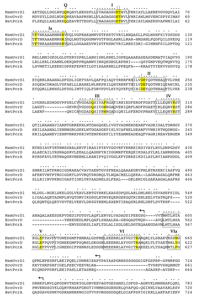
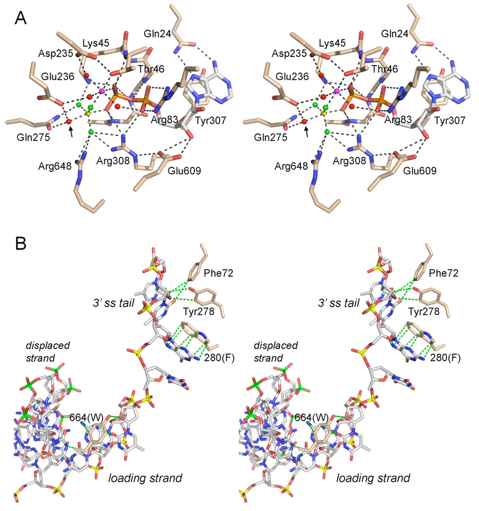
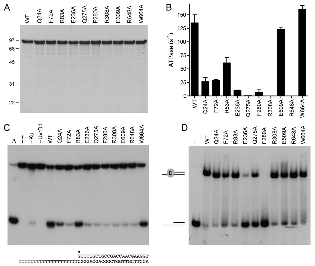
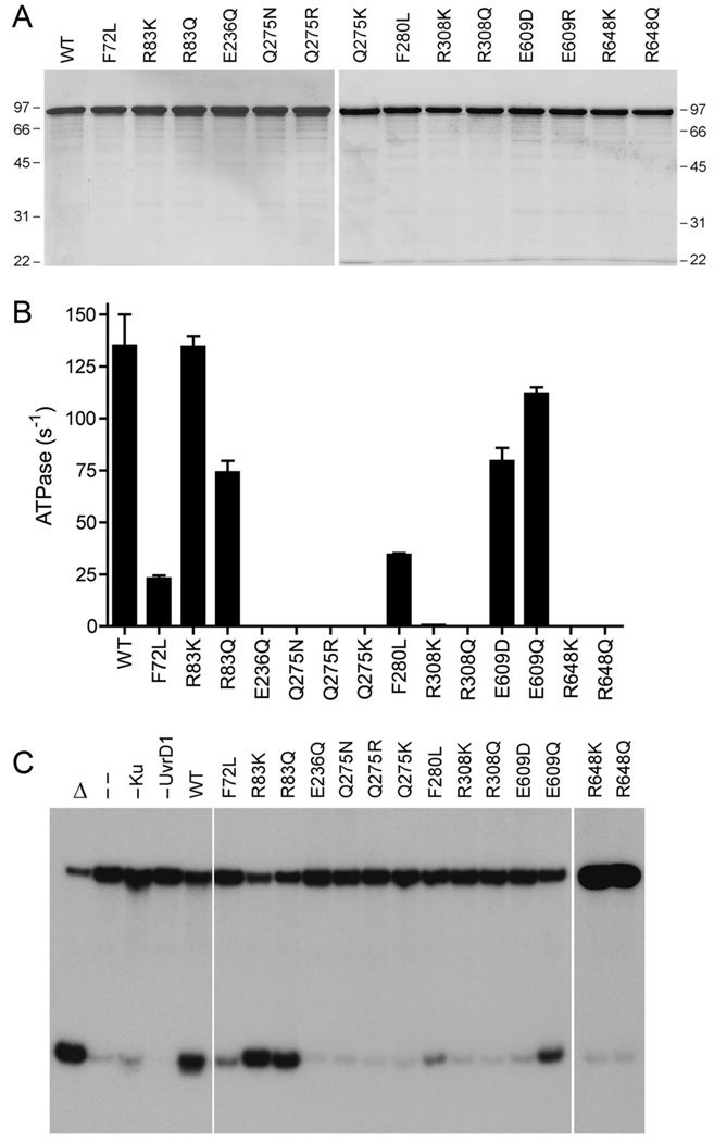
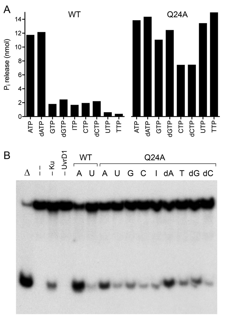
Mycobacterial UvrD1 is a DNA-dependent ATPase and a Ku-dependent 3' to 5' DNA helicase. The UvrD1 motor domain resembles that of the prototypal superfamily I helicases UvrD and PcrA. Here we performed a mutational analysis of UvrD1 guided by the crystal structure of a DNA-bound Escherichia coli UvrD-ADP-MgF(3) transition state mimetic. Alanine scanning and conservative substitutions identified amino acids essential for both ATP hydrolysis and duplex unwinding, including those implicated in phosphohydrolase chemistry via transition state stabilization (Arg308, Arg648, Gln275), divalent cation coordination (Glu236), or activation of the nucleophilic water (Glu236, Gln275). Other residues important for ATPase/helicase activity include Phe280 and Phe72, which interact with the DNA 3' single strand tail. ATP hydrolysis was uncoupled from duplex unwinding by mutations at Glu609 (in helicase motif V), which contacts the ATP ribose sugar. Introducing alanine in lieu of the adenine-binding "Q motif" glutamine (Gln24) relaxed the substrate specificity in NTP hydrolysis, e.g., eliciting a gain of function as a UTPase/TTPase, although the Q24A mutant still relied on ATP/dATP for duplex unwinding. Our studies highlight the role of the Q motif as a substrate filter and the contributions of adenosine-binding residues as couplers of NTP hydrolysis to motor activity. The Ku-binding function of UvrD1 lies within its C-terminal 270 amino acid segment. Here we found that deleting the 90 amino acid C-terminal domain, which is structurally uncharacterized, diminished DNA unwinding, without affecting ATP hydrolysis or binding to the DNA helicase substrate, apparently by affecting the strength of the UvrD1-Ku interaction.


Figures

Fig. 1
Fig. 1. Structural similarity between UvrD1, UvrD and PcrA
The amino acid sequence of M. smegmatis (Msm) UvrD1 (accession YP_889771) is aligned to the sequences of E. coli (Eco) UvrD (accession NP_418258) and B. stearothermophilus (Bst) PcrA (accession P56255). Positions of side chain identity/similarity in all three proteins are indicated by •. Gaps in the alignment are denoted by –. The ATPase/helicase motifs are denoted in boxes and named according to Lee and Yang (4). The conserved residues comprising the ATPase active site and DNA-binding interfaces of UvrD/PcrA that were subjected to alanine scanning in UvrD1 are highlighted in yellow. Essential constituents of UvrD1 ATPase motifs I (Lys45, Thr46) and II (Asp235) that were identified previously are indicated by |. The targeted UvrD1 residues, and their equivalents in UvrD and PcrA, are listed in Table I. The C-terminal truncations of UvrD1 are denoted by ↰.
Fig. 2
Fig. 2. ATPase active site and DNA contacts
(A) Stereo view of the phosphohydrolase active site of DNA-bound E. coli UvrD (PDB 2IS6) as a transition state mimetic in complex with Mg2+ (magenta sphere), ADP (stick model with gray carbons), and a trigonal planar MgF3 (depicted with the magnesium as a yellow sphere and the fluorines as green spheres). Waters are rendered as red spheres. The putative water nucleophile located apical to the leaving β-phosphate oxygen is indicated by an arrow. The atomic contacts of the conserved active site residues (stick models with beige carbons) are denoted by dashes lines. The residue numbers refer to the equivalent side chains in UvrD1 that were subjected to alanine scanning. (B) Stereo view of DNA contacts in the crystal structure of E. coli UvrD bound to a 3’ tailed duplex DNA (from PDB 2IS6). The 3’ tailed strand (the loading strand for initial helicase binding, on which the enzyme translocates 3’ to 5’) is depicted as a stick model with phosphorus atoms colored yellow. The complementary strand that forms the duplex segment (the displaced strand unwound by helicase translocation) is depicted with phosphorus atoms in green. Conserved aromatic residues that make base stacking and van der Waals contacts with the DNA (green dashed lines) are shown, with residue numbering referring to the equivalents in UvrD1.
Fig. 3
Fig. 3. Effects of alanine mutations on UvrD1 activities
(A) Aliquots (5 µg) of recombinant wild-type (WT) UvrD1 and the indicated Ala mutants were analyzed by SDS-PAGE. The Coomassie blue-stained gel is shown. The sizes (kDa) and positions of marker proteins are indicated on the left. (B) ATPase specific activity was determined as specified under Methods. Each datum is the average of two separate UvrD1 titration experiments; error bars denote the mean absolute error. (C) Helicase reactions were performed as described under Methods. Complete reaction mixtures contained 1 mM ATP, 50 nM 32P-labeled tailed duplex DNA substrate, 75 ng Ku and 100 ng of wild-type or mutant UvrD1 protein as specified. The products were analyzed by native PAGE and visualized by autoradiography. Reactions without added protein (lane --), with wild-type UvrD1 only (–Ku) or with Ku only (–UvrD1) were included as controls. A reaction lacking protein that was heat denatured prior to PAGE is shown in lane Δ. The 3’-tailed duplex helicase substrate is shown at bottom with the 5’ 32P-label denoted by •. (D) Binding of UvrD1 to the helicase substrate. Binding reactions were performed as described under Methods. Complete reaction mixtures contained 1 pmol 32P-labeled tailed duplex DNA substrate and 100 ng wild-type or mutant UvrD1 protein as specified. UvrD1 was omitted from the control reaction in lane –. The products were analyzed by native PAGE and visualized by autoradiography. The positions of the free DNA and the UvrD1-DNA complex are indicated on the left.
Fig. 4
Fig. 4. Effects of conservative mutations on UvrD1 activities
(A) Aliquots (3 µg) of recombinant wild-type (WT) UvrD1 and the indicated mutants were analyzed by SDS-PAGE. The Coomassie blue-stained gels are shown. The sizes (kDa) and positions of marker proteins are indicated. (B) ATPase specific activity was determined as specified under Methods. Each datum is the average of three separate UvrD1 titration experiments; error bars denote the standard deviation. (C) Helicase reactions were performed as described under Methods. Complete reaction mixtures contained 1 mM ATP, 50 nM 32P-labeled tailed duplex DNA substrate, 75 ng Ku and 100 ng of wild-type or mutant UvrD1 protein as specified. The products were analyzed by native PAGE and visualized by autoradiography. Reactions without added protein (lane --), with wild-type UvrD1 only (–Ku) or with Ku only (–UvrD1) were included as controls. A reaction lacking protein that was heat denatured prior to PAGE is shown in lane Δ.
Fig. 5
Fig. 5. Role of the Q motif (Gln24) in NTP substrate specifity
(A) Phosphohydrolase reaction mixtures contained 1 mM of the indicted ribonucleoside or deoxyribonucleoside triphosphate and either 50 ng wild-type UvrD1 or 200 ng of the Q24A mutant. The extents of phosphate production are shown. (B) Complete helicase reaction mixtures contained 50 nM 32P-labeled tailed duplex DNA substrate, 75 ng Ku, 100 ng of wild-type UvrD1 or Q24A, and 1 mM of the indicated NTP or dNTP. The products were analyzed by native PAGE and visualized by autoradiography. Reactions without added protein (lane --), with wild-type UvrD1 only (–Ku) or with Ku only (–UvrD1) were included as controls. A reaction mixture lacking protein that was heat denatured prior to PAGE is shown in lane Δ.
Fig. 6
Fig. 6. Effects of C-terminal deletions
(A) Aliquots (5 µg) of full-length UvrD1-(1–783) and the C-terminal truncation mutants UvrD1-(1–729), UvrD1-(1–693) and UvrD1-(1–595) were analyzed by SDS-PAGE. The Coomassie blue-stained gel is shown. The sizes (kDa) and positions of marker proteins are indicated on the left. (B) ATPase assays were performed as described in Methods. 32Pi release from 1 mM [γ32P]ATP is plotted as a function of input UvrD1 for each enzyme assayed. (C) Complete helicase reaction mixtures contained 1 mM ATP, 50 nM 32P-labeled tailed duplex DNA substrate, 75 ng Ku (where indicated by +) and 100 ng UvrD1 protein as specified. The products were analyzed by native PAGE and visualized by autoradiography. A reaction lacking protein that was heat-denatured prior to PAGE is shown in lane Δ. (D) DNA binding reaction mixtures contained 0.5 pmol 32P-labeled 3’-tailed DNA, 100 ng UvrD1-(1–783) or UvrD1-(1–693), and 75 ng Ku (where indicated by +). The free DNA and protein-DNA complexes (depicted on the right) were resolved by native PAGE and visualized by autoradiography.
Fig. 7
Fig. 7. Effect of 3’ tail length on helicase activity and DNA binding
(A) Helicase reaction mixtures contained 50 nM duplex DNA substrate with 3’-T20, 3’-T15, 3’-T10 or 3’-T5 tails, 1 mM ATP, 75 ng Ku, and 0, 25, 50 or 100 ng UvrD1. The products were analyzed by native PAGE and visualized by autoradiography. A control reaction lacking enzyme that was heat denatured prior to PAGE is shown in lane Δ. The 3’-T20 substrate is shown at bottom with the 5’ 32P-label denoted by •. (B) DNA binding reaction mixtures contained 0.5 pmol 32P-labeled 3’-tailed DNAs as specified, 100 ng UvrD1 (where indicated by +), and 75 ng Ku (where indicated by +). The products were analyzed by native gel electrophoresis. The free DNAs and various protein-DNA complexes (depicted at left and right) were visualized by autoradiography.

Similar articles

Cited by

References

    1. Singleton MR, Dillingham MS, Wigley DB. Structure and mechanism of helicases and nucleic acid translocases. Annu. Rev. Biochem. 2007;76:23–50. - PubMed
    1. Korolev S, Hsieh J, Gauss GH, Lohman TM, Waksman G. Major domain swiveling revealed by the crystal structures of complexes of E. coli Rep helicase bound to single-stranded DNA and ADP. Cell. 1997;90:635–647. - PubMed
    1. Velankar SS, Soultanas P, Dillingham MS, Subramanya HS, Wigley DB. Crystal structure of complexes of PcrA DNA helicase with a DNA substrate indicate an inchworm mechanism. Cell. 1999;87:75–84. - PubMed
    1. Lee JY, Yang W. UvrD helicase unwinds DNA one base pair at a time by a two-part power stroke. Cell. 2006;127:1349–1360. - PMC - PubMed
    1. Dillingham MS, Wigley DB, Webb MR. Demonstration of unidirectional single-stranded DNA translocation by PcrA helicase: measurement of step size and translocation speed. Biochemistry. 2000;39:205–212. - PubMed

Publication types

MeSH terms