. 2009 Apr 15;15(8):2723-30.
doi: 10.1158/1078-0432.CCR-08-1898. Epub 2009 Mar 24.

Expression of hepatocyte growth factor and its receptor met in Wilms' tumors and nephrogenic rests reflects their roles in kidney development

Affiliations

Expression of hepatocyte growth factor and its receptor met in Wilms' tumors and nephrogenic rests reflects their roles in kidney development

Raisa Vuononvirta et al. Clin Cancer Res. .

Abstract

Purpose: Hepatocyte growth factor (HGF) and its receptor Met are known to play diverse roles in both organogenesis and cancer. Wilms' tumor (WT) is a prototype for the link between abrogated development and neoplasia, with dysregulation of growth factor/receptor pathways playing key roles. Despite this, an understanding of the HGF/Met axis in the process is lacking.

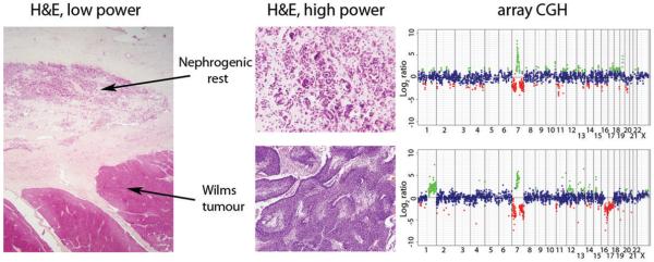
Experimental design: Observing copy number alterations at the loci for these genes in WTs and their precursor lesions nephrogenic rests, we examined protein expression by immunohistochemistry and investigated the effects of HGF on an in vitro model of kidney development.

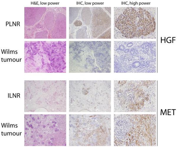
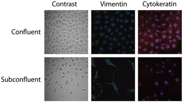
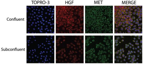
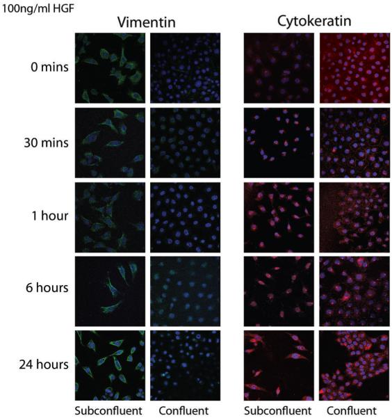
Results: HGF was preferentially expressed in the blastemal cells of nephrogenic rests but not WTs. Met expression was infrequent and restricted to well-differentiated epithelial cells and stroma in both lesions. In an independent cohort of favorable histology WTs on a tissue microarray, HGF was expressed in 15 of 193 (8%) cases and correlated with a predominance of epithelial cells, whereas Met expression was observed in 25 of 179 (14%) cases and was associated with stromal subtypes. In a mouse mesonephric cell line model, we observed Met expression in culture conditions reflecting both mesenchymal and epithelial differentiation, whereas HGF was up-regulated in association with acquisition of a more epithelial-like phenotype. This could be mimicked by exogenous exposure of mesenchymal-like cells to recombinant HGF.

Conclusions: These data show that the relatively infrequent expression of HGF and Met in WT tumorigenesis reflects their roles in nephrogenesis, particularly the mesenchymal-to-epithelial transition, rather than a dependence on oncogenic signaling pathways.


Figures

Figure 1
Figure 1. Array CGH of a nephrogenic rest and Wilms tumour from the same patient harbouring high level gain of 7q21
Haematoxylin and eosin stained sections (low power, original magnification ×12.5; high power, original magnification ×200) from a perilobar nephrogenic rest and associated blastemal type Wilms tumour alongside copy number profile by array CGH. Both lesions harbour a complex rearrangement on chromosome 7 leading to gain of 7q21 and concurrent loss of 7p and 7q22-qter. Additional alterations including gain of 1q and loss of 16q are present in the Wilms tumour. Genome plots show log2 ratios for each clone (x axis) plotted according to chromosomal location (y axis). The centromere is represented by a horizontal line. Points are coloured green and red to represent gains and losses, respectively.
Figure 2
Figure 2. Expression of HGF and Met in whole sections of nephrogenic rests and Wilms tumours
Photomicrographs demonstrating typical patterns of HGF and Met expression by immunohistochemistry in distinct nephrogenic rests and Wilms tumours from different patient samples. First row, a perilobar nephrogenic rest (PLNR) with strong, diffuse HGF staining in blastemal cells. Second row, a Wilms tumour demonstrating no immunoreactivity. Third row, an intralobar nephrogenic rest (ILNR) with strong Met expression in the stromal compartment. Fourth row, a Wilms tumour with strong stromal immunoreactivity. Low power, original magnification ×200; high power, original magnification ×400.
Figure 3
Figure 3. Mouse mesonephric M15 cells mimicking the mesenchymal to epithelial transition during nephrogenesis
M15 cells grown at different cell densities showing an epithelial morphology (phase contrast microscopy) and cytokeratin expression (red immunofluorescence) at confluence, and a mesenchymal phenotype (spindle shaped morphology and vimentin expression, green) when grown in subconfluent conditions. Nuclei were counterstained with TOPRO-3.
Figure 4
Figure 4. Expression of HGF and Met in M15 cells with epithelial and mesenchymal phenotypes
M15 cells grown at confluence and subconfluence stained by immunofluorescence for HGF (red, cytoplasm) and Met (green, cell membrane). Consistent staining is observed for Met, whilst an upregulation of HGF is seen in confluent conditions. Nuclei were counterstained with TOPRO-3. Merge indicates an integrated image of all three channels.
Figure 5
Figure 5. HGF drives the acquisition of an epithelial phenotype in subconfluent M15 cells
M15 cells grown in confluent and subconfluent conditions were treated with 100ng/ml recombinant HGF and stained for vimentin (green) and cytokeratin (red) by immunofluorescence. Nuclei were counterstained with TOPRO-3. An upregulation of cytokeratin was observed after 1 hour in the subconfluent cultures, demonstrating a distinct epithelial filament expression pattern at 24 hours.

References

    1. Horster MF, Braun GS, Huber SM. Embryonic renal epithelia: induction, nephrogenesis, and cell differentiation. Physiol Rev. 1999;79:1157–91. - PubMed
    1. Rivera MN, Haber DA. Wilms’ tumour: connecting tumorigenesis and organ development in the kidney. Nat Rev Cancer. 2005;5:699–712. - PubMed
    1. Schedl A. Renal abnormalities and their developmental origin. Nat Rev Genet. 2007;8:791–802. - PubMed
    1. Beckwith JB, Kiviat NB, Bonadio JF. Nephrogenic rests, nephroblastomatosis, and the pathogenesis of Wilms’ tumor. Pediatr Pathol. 1990;10:1–36. - PubMed
    1. Vuononvirta R, Sebire NJ, Dallosso AR, et al. Perilobar nephrogenic rests are non-obligate molecular genetic precursor lesions of IGF2-associated Wilms tumours. Clin Cancer Res. in press. - PMC - PubMed

Publication types

MeSH terms