Pontine reticular formation neurons: relationship of discharge to motor activity
- PMID: 193185
- PMCID: PMC9044325
- DOI: 10.1126/science.193185
Pontine reticular formation neurons: relationship of discharge to motor activity
Abstract
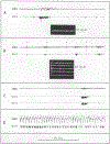
The discharge correlates of pontine reticular formation units were investigated in unrestrained cats. In agreement with previous investigations using immobilized preparations, we found that these cells had high rates of activity in rapid eye movement sleep, and responded in waking to somatic, auditory, and vestibular stimuli at short latencies, many having polysensory responses and exhibiting rapid "habituation." However, despite the sensory responses of these cells, most unit activity could not be explained by the presence of sensory stimuli. Intense firing occurred in association with specific movements. Units deprived of their adequate somatic, vestibular, and auditory stimuli showed undiminished discharge rates during motor activity. Discrete sensory stimuli evoked sustained unit firing only when they also evoked a motor response. We conclude that activity in pontine reticular formation neurons is more closely related to motor output than to sensory input.
Figures

References
-
- Amassian VE and Devito RV, J. Neurophysiol. 17, 575 (1954); - PubMed
- Bradley WE and Conway CJ, Exp. Neurol. 16, 237 (1966); - PubMed
- Pompeiano O and Barnes CD, J. Neurophysiol.’ 34, 709 (1971); - PubMed
- Glickstein M, Stein J, King RA, Science 178, 1110 (1972); - PubMed
- Motokizawa F,Exp. Neurol. 44, 135 (1974); - PubMed
- Peterson BW, Filion M, Felpel LP, Abzug C, Exp. Brain Res. 22, 335 (1975);
- Nagano S, Myers JA, Hall RD,Exp. Neurol. 49, 653 (1974); - PubMed
- Hornby JB and Rose JD, ibid. 51,363 (1976). - PubMed
-
- Scheibel M, Scheibel A, Mollica A, Moruzzi G,j. Neurophysiol. 18, 309 (1955); - PubMed
- Bach-Y-Rita P, Exp. Neurol. 9, 327 (1964); - PubMed
- Scheibel ME and Scheibel AB, Arch. Ital. Biol. 103, 279 (1965); - PubMed
- Segundo JP, Takenaka T, Encabo H, J. Neurophysiol 30,1221 (1967); - PubMed
- Peterson BW, Franck JI, Pitts NG, Daunton NG,ibid. 39, 564 (1976). - PubMed
-
- The pontine gigantocellular tegmental field as defined by Berman AL in The Brain Stem of the Cat (Univ. of Wisconsin Press, Madison, 1968) includes parts of the reticularis pontis oralis and caudalis nuclei described in earlier nomenclature.
-
- Responses of PRF units to noxious stimuli have been examined in acute and chronic studies including: Casey KL, Science 173, 77 (1971); - PubMed
- Barnes KL, Exp. Neurol. 50, 180 (1976); - PubMed
- Nyquist JK and Greenhoot JH, Brain Res. 70, 157 (1974); - PubMed
- Young DW and Gottschaldt KM, Exp. Neurol. 51,628 (1976); and - PubMed
- Sun CL and Gatipon GB, ibid. 52, 1 (1976). - PubMed
Publication types
MeSH terms
Grants and funding
LinkOut - more resources
Full Text Sources
Miscellaneous

