. 2009 Jul;19(7):743-55.
doi: 10.1093/glycob/cwp042. Epub 2009 Mar 24.

Inactivation of Mycobacterium tuberculosis mannosyltransferase pimB reduces the cell wall lipoarabinomannan and lipomannan content and increases the rate of bacterial-induced human macrophage cell death

Affiliations

Inactivation of Mycobacterium tuberculosis mannosyltransferase pimB reduces the cell wall lipoarabinomannan and lipomannan content and increases the rate of bacterial-induced human macrophage cell death

Jordi B Torrelles et al. Glycobiology. 2009 Jul.

Abstract

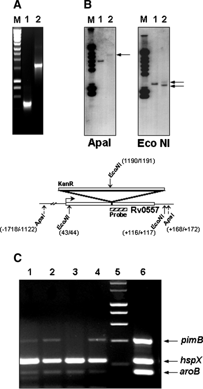
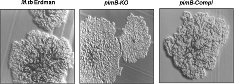
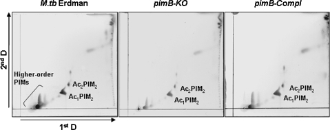
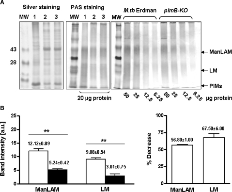
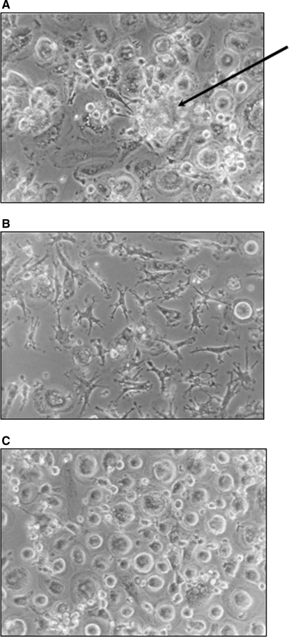
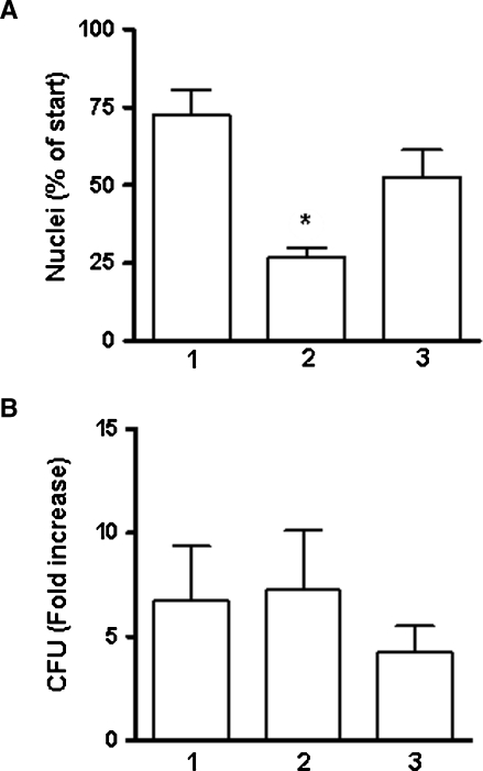
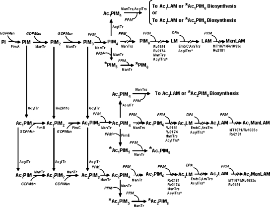
The Mycobacterium tuberculosis (M.tb) cell wall contains an important group of structurally related mannosylated lipoglycans called phosphatidyl-myo-inositol mannosides (PIMs), lipomannan (LM), and mannose-capped lipoarabinomannan (ManLAM), where the terminal alpha-[1-->2] mannosyl structures on higher order PIMs and ManLAM have been shown to engage C-type lectins such as the macrophage mannose receptor directing M.tb phagosome maturation arrest. An important gene described in the biosynthesis of these molecules is the mannosyltransferase pimB (Rv0557). Here, we disrupted pimB in a virulent strain of M.tb. We demonstrate that the inactivation of pimB in M.tb does not abolish the production of any of its cell wall mannosylated lipoglycans; however, it results in a quantitative decrease in the ManLAM and LM content without affecting higher order PIMs. This finding indicates gene redundancy or the possibility of an alternative biosynthetic pathway that may compensate for the PimB deficiency. Furthermore, infection of human macrophages by the pimB mutant leads to an alteration in macrophage phenotype concomitant with a significant increase in the rate of macrophage death.


Figures

Fig. 1
Fig. 1
Validation of the mutants used in this study. (A) PCR of mycobacterial genomic DNA demonstrating successful allelic exchange. Products of PCR reactions were separated on 1% agarose gel, stained with ethidium bromide, and visualized by UV exposure. The pimB PCR product (360 bp) in control M.tb Erdman DNA (lane 1). The pimB amplicon following allelic exchange was increased by the size of the aph contruct (1300 bp) to yield the expected product of 1660 bp (lane 2). M refers to DNA ladder. (B) Southern blot of mycobacterial ssDNA showing homologous recombination. Genomic DNA was enzyme-treated with ApaI (left panel) and EcoNI (right panel), and a Southern blot probed with a 32P-labeled fragment that spanned the KanR (aph) insertion site (see inset scheme). Predicted fragments were observed (arrows), M, DNA marker, lane 1, Erdman, lane 2 pimB-KO. (C) RT-PCR of RNA isolated from the pimB-KO demonstrates lack of RNA transcript for pimB mRNA and detection of hspX (encoding alpha crystallin homolog) and aroB (encoding dehydroquinate synthase) mRNAs, which serve as controls for RNA isolation and RT-PCR. Lanes: 1, Erdman; 2, PimB-Compl; 3, pimB-KO; 4, pimB-Overexp; 5, 200 bp ladder; 6, Erdman genomic DNA control. Controls for DNA contamination of RNA preparations demonstrated a lack of PCR product following amplification of cDNA without the addition of reverse transcriptase (data not shown).
Fig. 2
Fig. 2
Colony morphotypes showing alterations in the pimB-KO strain. The smooth morphology seen in the pimB-KO strain is in contrast to the colony morphology in the wild-type and complemented strains. Strains were grown on 7H11 agar (100× magnification).
Fig. 3
Fig. 3
Analysis of PIM profiles by 2D-TLC demonstrates that the product described for PimB, Ac1PIM2, is present in the pimB-KO strain. Lipids were extracted from the indicated strains and analyzed by 2D-TLC from samples based on equal amounts of protein. No differences in the PIM profiles were apparent, either at the PIM species level or in their acylation status. The individual species of PIMs observed are noted by abbreviations following the nomenclature AcxPIMy (PIM species with “x” (1 or 2) refers to the number of fatty acyl chains linked to either the core á(1→2)-linked Manp or the myo-inositol head group, and “y” represents the number of Manp residues). Shown is a representative experiment of six independent experiments.
Fig. 4
Fig. 4
Confirmation that pimB-KO produces Ac1PIM2 and Ac2PIM2 by MALDI-MS analysis. PIMs from the pimB-KO were extracted and purified by preparative-TLC (A) and subjected to MALDI-MS for identification (B). Band A was identified as m/z 1459.92, Ac1PIM2 with 2C16:0/TBST [M−H+2Na]+ and band B as m/z 1740.5, Ac2PIM2 with 2C16:0/2TBST [M−H+2Na]+.
Fig. 5
Fig. 5
ManLAM and LM amounts are decreased in the M.tb pimB-KO strain. (A) Up to 50 mg of cells (wet weight) were resuspended in 2 mL of PBS and disrupted by bead beating. The protein content was determined by BCA. Electrophoresis was performed using a 15% SDS–polyacrylamide gel with 20 μg of total protein for M.tb Erdman (lane 1), pimB-KO (lane 2), and pimB-Compl (lane 3) followed by staining with silver (left panel) to visualize the proteins or with PAS (central panel) to visualize the lipoglycans. In the right panel is shown a representative gel (n = 3 by duplicate) of M.tb Erdman and pimB-KO proteinase K-treated sonicates stained by PAS. A total of 50, 25, 12.5, or 6.25 μg of protein equivalents were loaded on the gel. ManLAM and LM are denoted by the arrows where there is a decrease in the ManLAM/LM content in pimB-KO when compared to M.tb Erdman which is most apparent at 6.25 μg protein equivalents (B) Densitometry analysis using the Image J program from NIH (http://rsb.info.nih.gov/ij/UT) at 6.25 μg protein equivalents revealed a reduction of 56.8 ± 1.0% for ManLAM and 67.5 ± 6.0% for LM. Data are presented as mean ± SEM, n = 6, **P < 0.005, Student's t-test.
Fig. 6
Fig. 6
Macrophage morphology observed following infection with M.tb demonstrates reduced homotypic adhesion in the pimB-KO strain when compared to the control M.tb Erdman strain. MDMs infected by M.tb Erdman strain (panel A) developed large aggregates over time (black arrow, panel A) when compared to pimB-KO (panel B), which had smaller aggregates. Macrophages were observed to have an elongated morphology with retracted cytoplasm. “Sham” uninfected monolayers (panel C) demonstrated no loss of cells, with macrophages maintaining a rounded, nonaggregate appearance at all times during the assay. Photomicrographs were taken by inverted phase microscopy at 200× at 72 h postinfection. Representative of three independent experiments in triplicate.
Fig. 7
Fig. 7
Macrophage viability decreases following infection with pimB-KO. (A) The percentage of macrophage nuclei remaining and (B) corresponding fold-increase in mycobacterial CFU. Quantification of macrophages was performed by isolation and counting of nuclei and corresponding CFUs from three independent donors, each of them measured in triplicate (mean ± SEM). Numbers reported are related to the value of CFUs or nuclei measured immediately following a 2 h infection with the M.tb strains. Time of culture following infection varied for individual donors in order to harvest cells when the monolayers were still at least 75% intact. Lane 1, M.tb Erdman; lane 2, pimB-KO; and lane 3, pimB-Compl. The only significant difference (*) in either percentage of nuclei or increase in CFU occurred in the case of the reduction in the numbers of nuclei in pimB-KO-infected macrophages, as compared to the control M.tb Erdman infection (2.7-fold reduction, *P < 0.05, Student's t-test).
Fig. 8
Fig. 8
A proposed scheme showing potential alternative pathways and redundancy in the biosynthesis pathway of PIMs, LM, and ManLAM. PimA, PimB, and PimC are three characterized manosyltransferases that use phosphatidyl-myo-inositol (PI) as a precursor lipid to synthesize lower order PIMs. PimE is implicated in the biosynthesis of higher order PIMs. MT1671/Rv1635c is the mannosyltransferase that adds the first mannose cap on M.tb ManLAM (Dinadayala et al. ; Appelmelk et al. 2008). The mannosyltransferase Rv2181 is described to have a dual function by adding α(1→2)-Manp to the ManLAM monocap to form the linear di- and trimannose caps and by adding α(1→2) branches to the mannan core of LM/LAM (Kaur et al. 2006, 2008). Rv2174 is proposed in building the linear α(1→6) mannan core (Kaur et al. ; Mishra et al. 2007). GDP-mannose (GDP-Man) (Besra et al. 1997) and polyprenol monophosphomannose (PPM) (Gurcha et al. ; Baulard et al. 2003) are the proposed mannose donors in this biosynthetic pathway. EmbC is described as an arabinosyltransferase/transporter using decaprenol monophosphate arabinose (DPA) as an arabinose donor (Zhang et al. ; Berg et al. 2005). There are still many unknown manosyltransferases (ManTrs), arabinosyltransferases (AraTrs), and acyltransferases (AcylTrs) involved in ManLAM biosynthesis. Alternative steps or redundancy present in this pathway may explain how it is possible to bypass a given enzyme. As depicted, in each step there is primarily one enzyme implicated. It is possible that in some steps (i.e., conversion of Ac1PIM1 to Ac1PIM2) more than one enzyme participates in the process emphasizing the plasticity that can exist in this pathway. Conversely, as shown for Rv2181, one enzyme may direct several steps reducing considerably the number of transferases implicated in this pathway. AcxPIMy represents nomenclature where it is implicit that PIMy already has two acyl groups located in its glycerol, “x” = 1 or 2 defines tri-acylated or tetra-acylated PIMy, and “y” denotes the number of mannoses. The same nomenclature is applied for AcxLM, AcXLAM, and AcxManLAM.*AcxPIMy components are produced from steps in the biosynthesis of ManLAM that deviate to alternatively synthesize*PIM6,*Ac1PIM6 or*Ac2PIM6 as final products. AcylTrans* stands for potential succinyl-, malyl- and/or lactyl-transferases.

Similar articles

Cited by

References

    1. Appelmelk BJ, den DJ, Driessen NN, Ummels R, Pak M, Nigou J, Larrouy-Maumus G, Gurcha SS, Movahedzadeh F, Geurtsen J, et al. The mannose cap of mycobacterial lipoarabinomannan does not dominate the Mycobacterium–host interaction. Cell Microbiol. 2008;10:930–944. - PubMed
    1. Astarie-Dequeker C, N’Diaye EN, Le Cabec V, Rittig MG, Prandi J, Maridonneau-Parini I. The mannose receptor mediates uptake of pathogenic and nonpathogenic mycobacteria and bypasses bactericidal responses in human macrophages. Infect Immun. 1999;67:469–477. - PMC - PubMed
    1. Barry CE III. Interpreting cell wall “virulence factors” of Mycobacterium tuberculosis. Trends Microbiol. 2001;9:237–241. - PubMed
    1. Baulard AR, Gurcha SS, Engohang-Ndong J, Gouffi K, Locht C, Besra GS. In vivo interaction between the polyprenol phosphate mannose synthase Ppm1 and the integral membrane protein Ppm2 from Mycobacterium smegmatis revealed by a bacterial two-hybrid system. J Biol Chem. 2003;278:2242–2248. - PubMed
    1. Berg S, Kaur D, Jackson M, Brennan PJ. The glycosyltransferases of Mycobacterium tuberculosis: Roles in the synthesis of arabinogalactan, lipoarabinomannan, and other glycoconjugates. Glycobiology. 2007;17:35–56R. - PubMed

Publication types

MeSH terms