. 2009 Apr 1;69(7):3105-13.
doi: 10.1158/0008-5472.CAN-08-3838. Epub 2009 Mar 24.

Triggering of Toll-like receptor 4 expressed on human head and neck squamous cell carcinoma promotes tumor development and protects the tumor from immune attack

Affiliations

Triggering of Toll-like receptor 4 expressed on human head and neck squamous cell carcinoma promotes tumor development and protects the tumor from immune attack

Miroslaw J Szczepanski et al. Cancer Res. .

Abstract

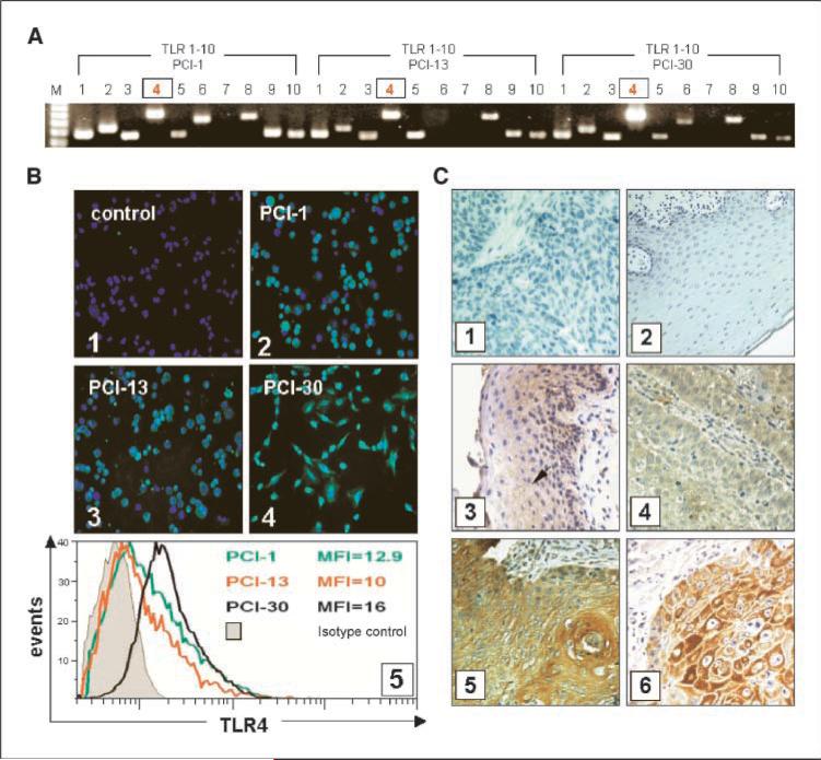
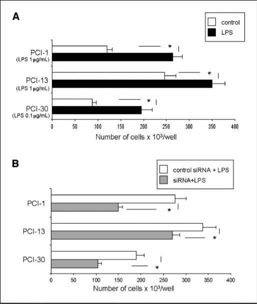
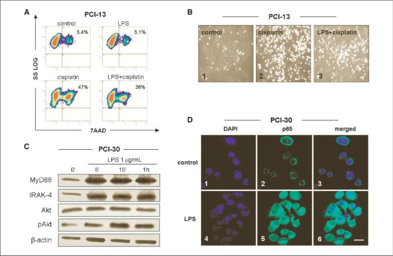
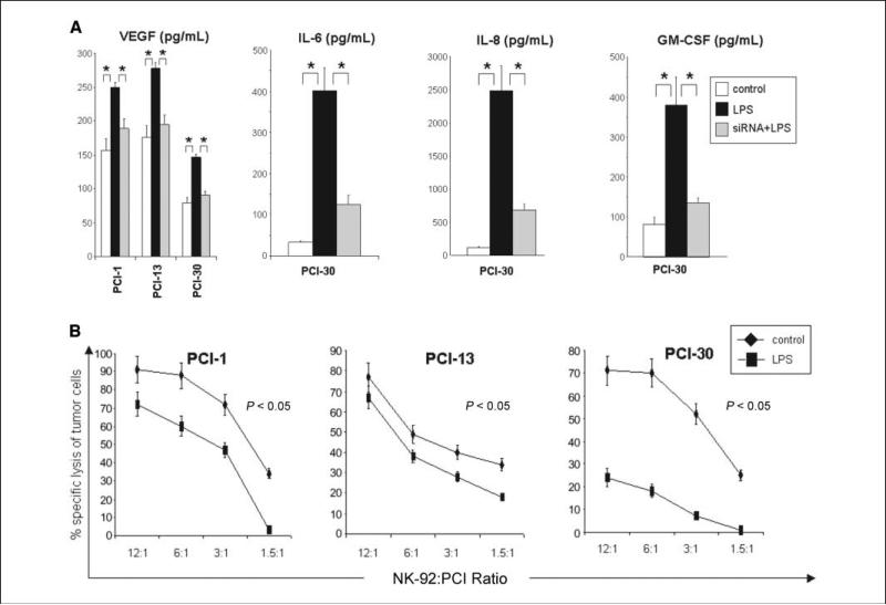
Toll-like receptors (TLR) expressed on inflammatory cells play a key role in host defense against pathogens, benefiting the host. TLR are also expressed on tumor cells. To evaluate the role of TLR in tumor cells, we investigated TLR4 signaling effects on human head and neck squamous cell carcinoma (HNSCC). Tumor tissues were obtained from 27 patients with laryngeal and 12 with oral cavity cancers. Normal mucosa was obtained from 10 patients with nonneoplastic disorders. Smears for bacteria were taken from all patients during surgery. TLR4 expression in tumors and HNSCC cell lines (PCI-1, PCI-13, and PCI-30) was detected by reverse transcription-PCR and immunohistochemistry. Cell growth, apoptosis, nuclear factor-kappaB (NF-kappaB) translocation, and MyD88 and IRAK-4 expression, as well as Akt phosphorylation were measured following tumor cell exposure to the TLR4 ligand lipopolysaccharide (LPS). Tumor cell sensitivity to NK-92-mediated lysis was evaluated in 4-hour (51)Cr-release assays. Cytokine levels in HNSCC supernatants were measured in Luminex-based assays. TLR4 was expressed in all tumors, HNSCC cell lines, and normal mucosa. The TLR4 expression intensity correlated with tumor grade. LPS binding to TLR4 on tumor cells enhanced proliferation, activated phosphatidylinositol 3-kinase/Akt pathway, up-regulated IRAK-4 expression, induced nuclear NF-kappaB translocation, and increased production (P<0.05) of interleukin (IL)-6, IL-8, vascular endothelial growth factor, and granulocyte macrophage colony-stimulating factor. TLR4 triggering protected tumor cells from lysis mediated by NK-92 cells. TLR4 ligation on tumor cells supports HNSCC progression.


Figures

Figure 1
Figure 1
TLR expression in tumors. TLR expression at the mRNA and protein levels in tumor cell lines, tumor, or normal mucosa. A, expression of TLR1 to TLR10 mRNA in the three HNSCC cell lines. B, expression of TLR4 protein in the three HNSCC cell lines. Magnification, ×400. 1, negative staining for TLR4 using isotype control IgG; 2, TLR4 in PCI-1; 3, TLR4 in PCI-13; 4, TLR4 in PCI-30 cells; 5, mean fluorescence intensity of TLR4 as determined by flow cytometry in the three cell lines. C, immunohistochemistry for TLR4 in tissue sections. Magnification, ×400. 1, isotype control in the tumor; 2, isotype control in the normal mucosa; 3, several TLR4+ cells in the normal mucosa (arrow); 4, a poorly differentiated HNSCC (G3) showing a weak positive reaction for TLR4; 5, a well-differentiated HNSCC (G1) showing strong positive reaction; 6, a representative HNSCC stained for cytokeratin.
Figure 2
Figure 2
Proliferation of tumor cells in response to LPS. A, PCI cell lines were incubated with 0.1 to 1.0 μg/mL of LPS. Each line was incubated with the LPS dose, which gives optimal tumor cell proliferation. Cells were counted on day 5 of culture. Control cultures contained no LPS. B, tumor cells were treated with TLR4-specific siRNA or with nonrelevant siRNA. A and B, columns, mean cell counts from three independent experiments; bars, SD. *, significant differences (P < 0.05) between experimental and control cultures.
Figure 3
Figure 3
Effects of LPS pretreatment on cisplatin-induced apoptosis and NF-κB activation in cancer cells. PCI cell lines were treated with 1 μg/mL LPS. A, LPS pretreatment protects tumor cells from apoptosis induced by cisplatin. Cell lines were first incubated ± LPS for 12 h and then with 10 μmol/L cisplatin for 24 h, stained with 7-AAD, and examined by flow cytometry. Results of a representative experiment of three performed are shown. B, morphologic changes in culture of PCI-13 cells incubated ± cisplatin or + LPS and cisplatin. Magnification, ×200. C, Western blots of PCI-30 cells incubated ± LPS show activation of the PI3K/Akt survival pathway. Increased levels of MyD88 and IRAK-4 in cells stimulated with LPS are accompanied by nuclear localization of the p65 subunit of NF-κB in LPS-treated PCI-30 cells. D, tumor cells plated overnight were stimulated with LPS (0.1 μg/mL) for 12 h and then stained as described in Materials and Methods. 1 to 3, control cells were untreated; 1, 3, 4, and 6, nuclei are stained blue; 2 and 5, the p65 subunit of NF-κB is stained green; 3, an overlay of 1 and 2; 6, an overlay of 4 and 5. At least 200 cells were randomly counted. Bar, 20 μm. Results are representative of three independent experiments.
Figure 4
Figure 4
Silencing of TLR4 using siRNA specific for TLR4. A, TLR4 was effectively silenced for at least 8 d as confirmed by RT-PCR and Western blot assays. B, the PI3K/Akt pathway in PCI-30 cells treated with control siRNA or siRNA specific for TLR4 and LPS. C, the p65 subunit binding activity or translocation of NF-κB into the nuclei in PCI-30 cells treated with control siRNA or siRNA specific for TLR4 and with LPS. *, significant increase or decrease of p65 binding activity at P < 0.05 in tumor cells stimulated with LPS or silenced for TLR4 expression, respectively.
Figure 5
Figure 5
Levels of cytokines in tumor cell supernatants and sensitivity of tumor to immune effector cells. A, supernatants were collected from tumor cells cultured at a density of 5 × 10⁵/mL after 48 h of stimulation and tested for levels of various cytokines in the presence or absence of siRNA and LPS as described in Materials and Methods. B, results of 51Cr-release assays with tumor cell targets stimulated with LPS (10 μg/mL). NK-92 cells are effector cells. The data are representative from three independent experiments with each tumor cell line used as targets.

References

    1. Medzhitov R, Preston-Hurlburt P, Janeway CA., Jr. A human homologue of the Drosophila Toll protein signals activation of adaptive immunity. Nature. 1997;388:394–7. - PubMed
    1. Stenzel W, Soltek S, Sanchez-Ruiz M, et al. Both TLR2 and TLR4 are required for the effective immune response in Staphylococcus aureus-induced experimental murine brain abscess. Am J Pathol. 2008;172:132–45. - PMC - PubMed
    1. Meier A, Alter G, Frahm N, et al. MyD88-dependent immune activation mediated by human immunodeficiency virus type 1-encoded Toll-like receptor ligands. J Virol. 2007;81:8180–91. - PMC - PubMed
    1. Akira S, Uematsu S, Takeuchi O. Pathogen recognition and innate immunity. Cell. 2006;124:783–801. - PubMed
    1. Elson G, Dunn-Siegrist I, Daubeuf B, Pugin J. Contribution of Toll-like receptors to the innate immune response to Gram-negative and Gram-positive bacteria. Blood. 2007;109:1574–83. - PubMed

Publication types

MeSH terms