. 2009 Apr 1;69(7):2878-86.
doi: 10.1158/0008-5472.CAN-08-2857. Epub 2009 Mar 24.

Caveolin-1 regulates the antagonistic pleiotropic properties of cellular senescence through a novel Mdm2/p53-mediated pathway

Affiliations

Caveolin-1 regulates the antagonistic pleiotropic properties of cellular senescence through a novel Mdm2/p53-mediated pathway

Janine N Bartholomew et al. Cancer Res. .

Abstract

We show that caveolin-1 is a novel binding protein for Mdm2. After oxidative stress, caveolin-1 sequesters Mdm2 away from p53, leading to stabilization of p53 and up-regulation of p21(Waf1/Cip1) in human fibroblasts. Expression of a peptide corresponding to the Mdm2 binding domain of caveolin-1 is sufficient to up-regulate p53 and p21(Waf1/Cip1) protein expression and induce premature senescence. Oxidative stress-induced activation of the p53/p21(Waf1/Cip1) pathway and induction of premature senescence are compromised in caveolin-1 null mouse embryonic fibroblasts (MEF). We also show that reintroduction of caveolin-1 in oncogenic Ras (Ras(G12V))-transformed fibroblasts, which express residual levels of caveolin-1, is sufficient to promote cellular senescence. Moreover, caveolin-1 expression in MEFs is required for senescent fibroblast-induced stimulation of cell growth and tumorigenesis of both Ras(G12V)-transformed fibroblasts and MDA-MB-231 breast cancer epithelial cells both in vitro and in vivo. Thus, our results propose caveolin-1 as a key mediator of the antagonistic pleiotropic properties of cellular senescence.


Figures
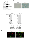
Figure 1
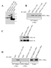
Figure 1. Sequestration of Mdm2 by caveolin-1 upon oxidant stimulation
(A) Immunoblotting. WI-38 cells were treated with subcytotoxic concentrations of hydrogen peroxide for 2 hours. Cells were washed and cultured in normal medium for 24 hours. Cell lysates were matched for total protein content by BCA protein assay and subjected to immunoblotting analysis using antibody probes against p53, p21WAF1/CIP1, and caveolin-1 (Cav-1). (B) Senescence-Associated (SA) β-galactosidase activity assay. WI-38 cells were treated as in (A). Cells were then subjected to senescence-associated β-galactosidase activity assay after 7 days of recovery following the H2O2 treatment using the Senescence-Associated β-galactosidase Staining Kit (Cell Signaling). A representative field is shown. (C) Immunoprecipitation assay. WI-38 human diploid fibroblasts were treated with 450 µM H2O2 for 2 hours. After twenty four hours, cell lysates were immunoprecipitated with a monoclonal antibody probe specific for Mdm2 and immunoprecipitates subjected to immunoblotting analysis with anti-caveolin-1 pAb. Total caveolin-1 and Mdm2 expression are shown in the lower panels. Immunoprecipitation with a monoclonal antibody probe specific for caveolin-3, a caveolin-1 homolog that is not expressed in fibroblasts, was used as negative control. (D) Immunofluorescence analysis. Wi-38 cells were treated as in (A). After twenty four hours of recovery following the H2O2 treatment, cells were double stained with anti-caveolin-1 pAb and anti-Mdm2 mAb. Untreated cells (-H2O2) were used as controls. The expression of these proteins was detected using fluorescent secondary antibodies. Data were collected sequentially in different channels. Representative images were taken using a 20X objective.
Figure 2
Figure 2. Identification of the Mdm2-binding domain of caveolin-1 and caveolin-1-binding motif of Mdm2
(A) and (B) In vitro reconstitution of Mdm2 binding to Caveolin-1. NIH 3T3 cells were transiently transfected with the cDNA encoding Mdm2, containing the c-myc tag at its C-terminus. Cell lysates were incubated with affinity-purified GST alone or GST fused to a series of caveolin-1 deletion mutants (Full-length (FL), residues 1–101, and residues 82–101) immobilized on glutathione-agarose beads (A). The beads were then separated by SDS-PAGE and subjected to Western blot analysis with anti-c-myc monoclonal antibody to detect Mdm2 binding (B). (C) Generation and expression of Φ→A Mdm2 mutant. NIH 3T3 cells were transiently transfected with wild type Mdm2 or a mutated form of Mdm2 in which the aromatic residues of the putative caveolin-1 binding motif were substituted with alanines. Transfection with the expression vector alone was used as a control. Forty eight hours after transfection, cell lysates were subjected to immunoblotting analysis using either anti-Mdm2 or anti-c-myc mAbs. (D) In vitro reconstitution of Φ→A Mdm2 mutant binding to the caveolin-1 scaffolding domain. The experiments were performed as described in (B) with the exception that Φ→A Mdm2-c-Myc mutant was transfected in addition to wild type Mdm2-c-Myc and cell lysates were incubated with only affinity-purified GST alone or GST-caveolin-1 82-101.
Figure 3
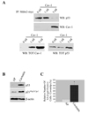
Figure 3. Caveolin-1 expression prevents Mdm2 binding to p53. Activation of the p53/p21Waf1/Cip1/senescence pathway by the caveolin-1 scaffolding domain
(A) Immunoprecipitation assay. NIH 3T3 fibroblasts were co-transfected with a c-myc-tagged Mdm2-expressing vector and increasing concentrations of a caveolin-1-expressing vector. Twenty four hours after transfection, cells were treated with 150 µM H2O2 for 2 hours. After twenty four hours, cell lysates were immunoprecipitated with a monoclonal antibody probe specific for c-myc and immunoprecipitates subjected to immunoblotting analysis with anti-caveolin-1 and anti-p53 pAbs. Total caveolin-1 and p53 expression are shown in the lower panels. (B) and (C) Immunoblotting and Senescence-associated (SA) β-galactosidase activity assay. WI-38 cells were incubated with either cavtratin (10µM) or the control peptide antennapedia (AP) (10µM) for either 24 hours (B) or 7 days (C). In (B), cell lysates were subjected to immunoblotting analysis using antibody probes against p53 (pAb) and p21Waf1/Cip1 (mAb). Immunoblotting with anti-β-actin mAb was performed to show equal loading. In (C), cells were subjected to senescence-associated β-galactosidase activity assay. Quantification of SA-β-gal-positive cells is shown. Values represent means ± SEM. *P<0.001.
Figure 4
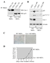
Figure 4. Activation of the p53/p21Waf1/Cip1 pathway and induction of premature senescence by oxidative stress are inhibited in caveolin-1 null MEFs
(A) Immunoprecipitation assay. Mouse embryonic fibroblasts were derived from wild type (WT MEFs) and caveolin-1 null (Cav-1 KO MEFs) mice. Cells were treated with 150 µM H2O2 for 2 hours, washed, and cultured in normal medium for 12 and 48 hours. Cell lysates were then immunoprecipitated with a monoclonal antibody probe specific for Mdm2 bound to agarose beads and immunoprecipitates subjected to immunoblotting analysis with anti-caveolin-1 and anti-p53 pAbs. (B) Immunoblotting. Wild type (WT MEFs) and caveolin-1 null (Cav-1 MEFs) were treated with 150 µM H2O2 for 2 hours, washed, and cultured in normal medium for 48 hours. Untreated cells were used as controls. Cell lysates were then subjected to immunoblotting analysis using antibody probes against p53 (pAb), p21Waf1/Cip1 (mAb), and caveolin-1 (pAb). Immunoblotting with anti-β-actin mAb was performed to show equal loading. (C) and (D) Senescence-Associated (SA) β-galactosidase activity assay. Wild type (WT MEFs) and caveolin-1 null (Cav-1 KO) MEFs were treated with 150 µM hydrogen peroxide for 2 hours. Cells were washed and cultured in normal medium for different periods of time (3, 5, 6, 8, and 10 days). Cells were then subjected to senescence-associated β-galactosidase activity assay. A representative field of the staining 10 days after oxidative stress is shown in (C). Quantitation of the SA-β-galactosidase activity assay is shown in (D).
Figure 5
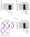
Figure 5. Caveolin-1 induces senescence of cancer cells and mediates senescent fibroblast-induced proliferation of cancer cells in vitro and in vivo
(A) Senescence-Associated (SA) β-galactosidase activity assay. Caveolin-1-and pCAGGS-expressing cells were derived as described in Supplementary Figure 5A. Cells were then subjected to senescence-associated β-galactosidase activity assay. Quantitation of the SA-β-galactosidase activity assay is shown. Values represent means ±SEM. *P<0.001. (B). Co-culture studies using RasG12V-transformed NIH 3T3 cells. RasG12V-transformed NIH 3T3 cells were cultured alone or with either wild type or caveolin-1 null MEFs as described in Materials and Methods. Cells were then stained with anti-Ki67 mAb. Ki67 staining was detected using fluorescent secondary antibodies. Quantification of Ki67 staining is shown. Values represent means ± SEM. *, #P<0.001. (C) Co-culture studies using MDA-MB-231 cells. MDA-MB-231 cells were cultured alone or with either wild type or caveolin-1 null MEFs as described in Materials and Methods. Colony formation was then detected by staining the cells with crystal violet (A representative image is shown in (C); quantification of the staining is represented in Supplementary Figure 5F). (D) Secreted factors studies. RasG12V-transformed NIH 3T3 cells were grown with secreted factors from hydrogen peroxide-treated wild type and caveolin-1 null MEFs as described in Materials and Methods. Cells were then stained with anti-Ki67 mAb. Ki67 staining was detected using fluorescent secondary antibodies. Quantification of Ki67 staining is shown. Values represent means ± SEM. *, #P<0.001.
Figure 6
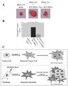
Figure 6. Caveolin-1 mediates senescent fibroblast-induced tumorigenesis in vivo
(A) and (B) Tumor xenografts studies in nude mice. MDA-231 cells were subcutaneously injected alone or with either hydrogen peroxide-treated wild type or caveolin-1 null MEFs into the dorsal flap of nude (nu / nu) mice. Mice were sacrificed four weeks later and tumor size measured. Representative images are shown in (A); quantification of tumor size is shown in (B). Values in (B) represent means ± SEM. *, #P<0.001. (C) Schematic diagram summarizing the role of caveolin-1 as a key mediator of the antagonistic pleiotropic properties of cellular senescence. Anti-tumorigenic effect of caveolin-1-mediated cellular senescence: by inducing cellular senescence of cancer cells, including breast cancer epithelial cells, caveolin-1 expression inhibits cancer cell growth. Pro-tumorigenic effect of caveolin-1-mediated cellular senescence: caveolin-1 indirectly promotes tumor cell growth by inducing senescence of fibroblasts, which release secreted factors that stimulate proliferation of cancer cells.

Similar articles

Cited by

References

    1. Williams GC. Pleiotropy, natural selection, and the evolution of senescence. Evolution. 1957;11:398–411.
    1. Campisi J. Senescent cells, tumor suppression, and organismal aging: good citizens, bad neighbors. Cell. 2005;120:513–522. - PubMed
    1. Campisi J. Cellular senescence as a tumor-suppressor mechanism. Trends Cell Biol. 2001;11:S27–S31. - PubMed
    1. Linskens MH, Feng J, Andrews WH, et al. Cataloging altered gene expression in young and senescent cells using enhanced differential display. Nucleic Acids Res. 1995;23:3244–3251. - PMC - PubMed
    1. Millis AJ, Hoyle M, McCue HM, Martini H. Differential expression of metalloproteinase and tissue inhibitor of metalloproteinase genes in aged human fibroblasts. Exp Cell Res. 1992;201:373–379. - PubMed

Publication types

Substances