. 2009;4(3):e4982.
doi: 10.1371/journal.pone.0004982. Epub 2009 Mar 25.

Virulent clones of Klebsiella pneumoniae: identification and evolutionary scenario based on genomic and phenotypic characterization

Affiliations

Virulent clones of Klebsiella pneumoniae: identification and evolutionary scenario based on genomic and phenotypic characterization

Sylvain Brisse et al. PLoS One. 2009.

Abstract

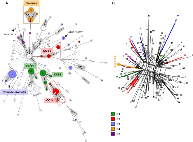
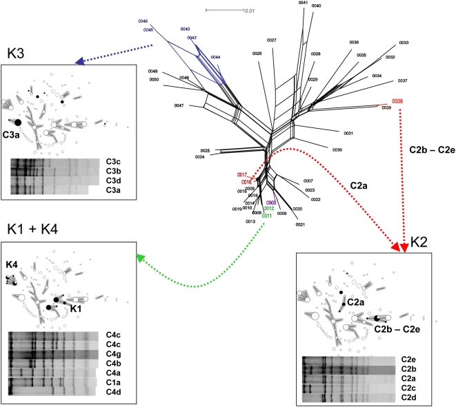
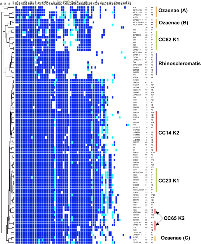
Klebsiella pneumoniae is found in the environment and as a harmless commensal, but is also a frequent nosocomial pathogen (causing urinary, respiratory and blood infections) and the agent of specific human infections including Friedländer's pneumonia, rhinoscleroma and the emerging disease pyogenic liver abscess (PLA). The identification and precise definition of virulent clones, i.e. groups of strains with a single ancestor that are associated with particular infections, is critical to understand the evolution of pathogenicity from commensalism and for a better control of infections. We analyzed 235 K. pneumoniae isolates of diverse environmental and clinical origins by multilocus sequence typing, virulence gene content, biochemical and capsular profiling and virulence to mice. Phylogenetic analysis of housekeeping genes clearly defined clones that differ sharply by their clinical source and biological features. First, two clones comprising isolates of capsular type K1, clone CC23(K1) and clone CC82(K1), were strongly associated with PLA and respiratory infection, respectively. Second, only one of the two major disclosed K2 clones was highly virulent to mice. Third, strains associated with the human infections ozena and rhinoscleroma each corresponded to one monomorphic clone. Therefore, K. pneumoniae subsp. ozaenae and K. pneumoniae subsp. rhinoscleromatis should be regarded as virulent clones derived from K. pneumoniae. The lack of strict association of virulent capsular types with clones was explained by horizontal transfer of the cps operon, responsible for the synthesis of the capsular polysaccharide. Finally, the reduction of metabolic versatility observed in clones Rhinoscleromatis, Ozaenae and CC82(K1) indicates an evolutionary process of specialization to a pathogenic lifestyle. In contrast, clone CC23(K1) remains metabolically versatile, suggesting recent acquisition of invasive potential. In conclusion, our results reveal the existence of important virulent clones associated with specific infections and provide an evolutionary framework for research into the links between clones, virulence and other genomic features in K. pneumoniae.


Conflict of interest statement

Competing Interests: The authors have declared that no competing interests exist.

Figures

Figure 1
Figure 1. Clonal diversity and relationships among 235 Klebsiella pneumoniae isolates.
A. Minimum spanning tree (MStree) analysis of multilocus sequence typing (MLST) data for 235 K. pneumoniae isolates, representing 117 sequence types (STs). Isolates of capsular serotypes K1 to K5 are colored according to serotype. Each circle corresponds to a sequence type (ST); ST number is given inside each circle. Grey zones surround STs that belong to the same clonal complex (CC), which is named according to the central ST (the likely founder of the CC). CC65-K2 is delimited by the red triangle (see text). The lines between STs indicate inferred phylogenetic relationships and are represented as bold, plain, discontinuous and light discontinuous depending on the number of allelic mismatches between profiles (1, 2, 3 and 4 or more, respectively); note that discontinuous links are only indicative, as several alternative links with equal weight may exist. The STs of reference genome strain MGH78578 (ST38) and of the type strain ATCC 13883T (ST3) are indicated. B. Split decomposition analysis of concatenated sequences of the seven genes. Numbers at the tip of branches are ST numbers. Note the bushy network structure indicative of pervasive homologous recombination. Branches were colored for the clones that are highlighted on panel A. Note the distribution into unrelated branches of strains with a given capsular (K) type.
Figure 2
Figure 2. Distribution of related capsular operon regions in unrelated clones.
The splits tree represents the relationships among gnd alleles as obtained after split decomposition analysis. The distribution of the gnd alleles found in isolates and reference strains of capsular serotypes K1 to K4 is indicated by black coloration of sequence types (STs) in the MStree of the corresponding insets. Below the MStree displays are represented the C-pattern of the corresponding isolates. Note that similar or identical gnd and C-patterns are distributed in unrelated STs.
Figure 3
Figure 3. Biotype profiling of K. pneumoniae clones.
Cluster analysis (simple matching coefficient) of K. pneumoniae isolates and reference strains based on metabolic profiles as assessed by biotype-100 strips. Codes above the column correspond to substrate code (Table S3). A blue square means the strain grew on the corresponding substrate as sole carbon source. Dark blue, growth was observed after two days; light blue, growth observed after four days. Note the strong homogeneity of biotype-100 profiles within clones. Three clones (Ozaenae, Rhinoscleromatis and CC82 K1) have lost the ability to utilize a number of substrates, including some common substrates between the three clones (see text). Note that three tests measure coloration, not growth: hydroxyquinoline-beta-glururonide (black color), tryptophane (brown color: hydrolysis into indole-pyruvic acid) and histidine (red color). The following substrates were utilized by all assayed strains: D-Glucose, D-fructose, D-trehalose, D-Melibiose, D-Raffinose, Maltotriose, Maltose, D-Cellobiose, 1-O-Methyl-B-D-glucoside, D-Arabitol, Glycerol, Adonitol, N-Acetyl-D-glucosamine, D-Gluconate, L-Alanine and L-Serine. The following substrates were always negative: hydroxyquinoline-beta-glucuronide, D-Lyxose, i-Erythritol, 3-O-Methyl-D-glucose, Tricarballylate, Tryptophan, Gentisate, 3-Hydroxybenzoate, 3-Phenylpropionate, Trigonelline, Betaine, Caprylate, Tryptamine, Itaconate, Propionate, 2-Ketoglutarate.

Similar articles

Cited by

References

    1. Ørskov I. The genus Klebsiella (Medical aspects). In: Starr MP, Stolp H, Truper HG, Balows A, Schlegel HG, editors. The Prokaryotes: a handbook on habitats, isolation, and identification of bacteria. Berlin: Springer-Verlag; 1981. pp. 1160–1165.
    1. Seidler RJ. The genus Klebsiella (Nonmedical aspects). In: Starr MP, Stolp H, Truper HG, Balows A, Schlegel HG, editors. The Prokaryotes: a handbook on habitats, isolation, and identification of bacteria. Berlin: Springer-Verlag; 1981. pp. 1166–1172.
    1. Brisse S, Grimont F, Grimont PAD. The genus Klebsiella. In: Dworkin M, Falkow S, Rosenberg E, Schleifer K-H, Stackebrandt E, editors. The Prokaryotes · A Handbook on the Biology of Bacteria. 3rd edition ed. New York: Springer; 2006.
    1. Podschun R, Ullmann U. Klebsiella spp. as nosocomial pathogens: epidemiology, taxonomy, typing methods, and pathogenicity factors. Clin Microbiol Rev. 1998;11:589–603. - PMC - PubMed
    1. Paterson DL, Ko WC, Von Gottberg A, Mohapatra S, Casellas JM, et al. International prospective study of Klebsiella pneumoniae bacteremia: implications of extended-spectrum beta-lactamase production in nosocomial Infections. Ann Intern Med. 2004;140:26–32. - PubMed

Publication types