. 2009;4(3):e4942.
doi: 10.1371/journal.pone.0004942. Epub 2009 Mar 25.

Dendritic cell-derived exosomes promote natural killer cell activation and proliferation: a role for NKG2D ligands and IL-15Ralpha

Affiliations

Dendritic cell-derived exosomes promote natural killer cell activation and proliferation: a role for NKG2D ligands and IL-15Ralpha

Sophie Viaud et al. PLoS One. 2009.

Abstract

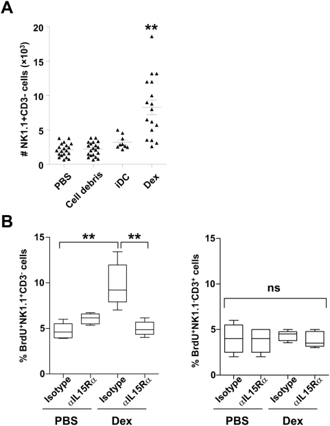
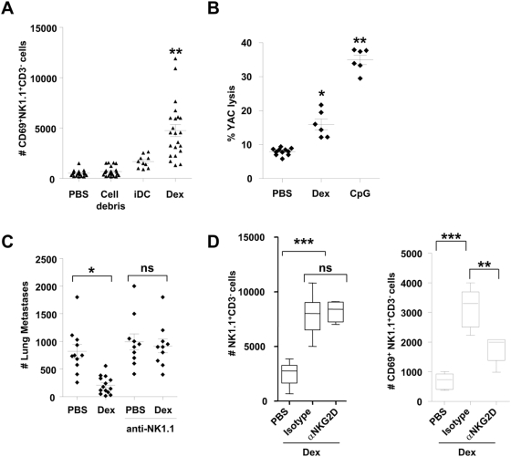
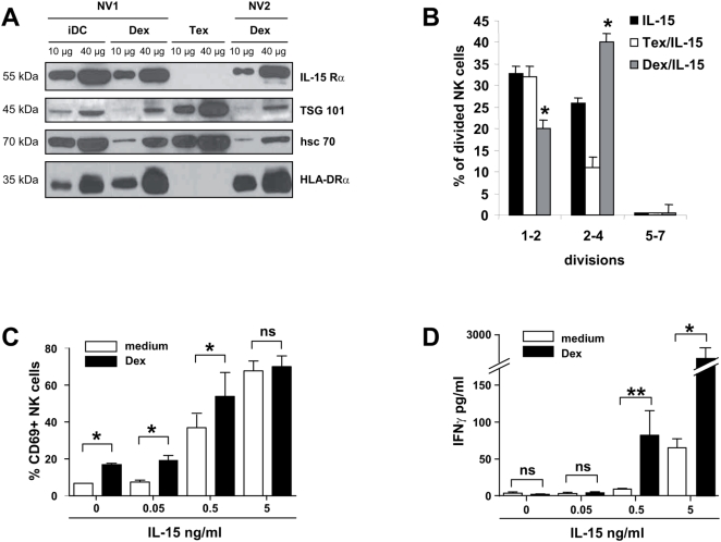
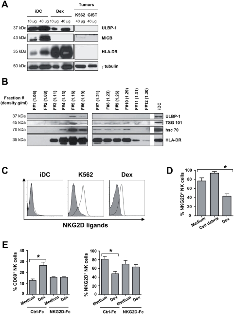
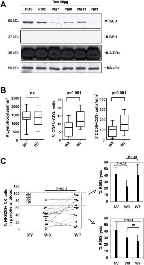
Dendritic cell (DC) derived-exosomes (Dex) are nanomeric vesicles harboring functional MHC/peptide complexes promoting T cell-dependent tumor rejection. In the first Phase I trial using peptide-pulsed Dex, the observation of clinical regressions in the absence of T cell responses prompted the search for alternate effector mechanisms. Mouse studies unraveled the bioactivity of Dex on NK cells. Indeed, Dex promoted an IL-15Ralpha- and NKG2D-dependent NK cell proliferation and activation respectively, resulting in anti-metastatic effects mediated by NK1.1(+) cells. In humans, Dex express functional IL-15Ralpha which allow proliferation and IFNgamma secretion by NK cells. In contrast to immature DC, human Dex harbor NKG2D ligands on their surface leading to a direct engagement of NKG2D and NK cell activation ex vivo. In our phase I clinical trial, we highlight the capacity of Dex based-vaccines to restore the number and NKG2D-dependent function of NK cells in 7/14 patients. Altogether, these data provide a mechanistic explanation on how Dex may stimulate non MHC restricted-anti-tumor effectors and induce tumor regression in vivo.


Conflict of interest statement

Competing Interests: The authors have declared that no competing interests exist.

Figures

Figure 1
Figure 1. Mouse Dex promoted NK cell proliferation: a role for IL-15Rα.
A. Dex induce NK cell influx in draining lymph nodes. Enumeration of CD3 NK1.1+ cells (NK cells) in the draining lymph node following intradermal inoculation of 10 µg of mouse Dex, or 3×10⁵ immature DC (iDC), or 10 µg of irrelevant pelleted proteins (cell debris) or PBS. B. NK cells enter cell cycle following Dex inoculation. Proportion of BrdU+ CD3 NK1.1+ cells (NK cells) (left panel) or BrdU+ CD3+ NK1.1 cells (T cells) (right panel) in the draining lymph node following intradermal inoculation of 10 µg of PBS or mouse Dex in the presence of anti-IL-15Rα blocking mAb (αIL-15Rα) or isotype control mAb (Isotype). The graphs depict the means of absolute numbers or percentages±SEM of the data from 4 pooled experiments.* p<0.05, ** p<0.01 and ns: “non significant”.
Figure 2
Figure 2. Mouse Dex promoted NK cell activation: a role for NKG2D.
A. Dex induced NK cell activation in the draining lymph node. Absolute numbers of CD3 NK1.1+ CD69+ cells in the draining lymph node following inoculation of 10 µg of mouse Dex, or 3×10⁵ iDC or 10 µg of irrelevant pelleted proteins (cell debris) or PBS. Each dot represents the result in one mouse. B. Dex stimulated splenic NK cytotoxicity. Killing assays on splenocytes against YAC-1 targets at 200∶1 and 50∶1 (not shown) after intradermal inoculations of 10 µg of Dex every other week for 8 weeks and sacrifice 48 hrs after the last immunization, or 24 hrs after a single subcutaneous injection of 10 µg of CpG ODN. C. Therapy with unpulsed Dex reduced number of metastases. Intradermal inoculation of 20 µg of exosomal proteins or PBS 5 days after intravenous injection of 3.105 B16F10 tumor cells. Depletion of NK cells was achieved using 3 administrations of anti-NK1.1 mAbs or isotype control mAbs. Mice were sacrificed on day 10 for enumeration of lung metastases. D. A role for NKG2D in Dex-mediated NK cell triggering. Enumeration of CD3NK1.1+ cells (left panel) and CD3 NK1.1+CD69+ cells (right panel) in the draining lymph node following intradermal inoculation of PBS or 10 µg of mouse Dex in the presence of anti-NKG2D blocking mAb (αNKG2D) or control isotype mAb. The graphs depict the data of 4 pooled experiments (2 for B.). Means and SEM are shown. * p<0.05, ** p<0.01, *** p<0.001 and ns: “non significant”.
Figure 3
Figure 3. Human Dex harbour functional IL-15Rα and synergize with IL-15 for NK cell proliferation in vitro and IFNγ production in vitro.
A. Immunoblotting of IL-15Rα from Dex and DC lysates. Western Blot analysis on 10–40 µg of protein lysates obtained from immature DC (iDC), Dex or Tex (exosomes from Mel888 melanoma cell line) using anti-IL-15Rα mAb. Positive controls included anti-HLA-DRα, -TSG 101 and -HSC 70 Abs. Representative immunoblots of two normal volunteers are depicted (NV1 and NV2). Molecular weights are indicated on the left lane. B. Proliferative effects of recombinant IL-15 and Dex on NK cells. CFSE-labeled NK cells were cultured with or without 10 µg autologous Dex or allogenic Tex in complete medium containing 0.5 ng/ml of human recombinant IL-15. At day 6 of culture, NK cell proliferation was determined by flow cytometry and the number of divisions were counted and depicted. A representative experiment out of two is shown. C–D. Synergistic effects between Dex and recombinant IL-15 for NK cell triggering. NV's PBL were cultured without (white histograms) or with (black histograms) 10 µg autologous Dex and increasing concentrations of human recombinant IL-15. NK (CD56+ CD3) cells were then analysed for CD69 expression by flow cytometry (C) or supernatants were harvested to measure IFNγ levels in EIA (D). The graphs depict means±SEM of % of CD69 expressing NK cells in 3 experiments (C) or IFNγ concentrations in 4 experiments (D). * p<0.05, ** p<0.01 and ns: non significant.
Figure 4
Figure 4. Human Dex harbour functional NKG2D ligands.
A–B. Immunoblot detection of NKG2D ligands on Dex. (A) 10–40 µg of protein lysates from immature DC (iDC), Dex or tumor cells (K562, GIST) were assayed in western blot analyses using anti-ULBP-1 and MICB Abs on whole protein lysates (A) (this result was similar in 6 Dex preparations from 6 different healthy donors) or on each fraction of distinct density (B) following ultracentrifugation of 400 µg of Dex proteins on a continuous density gradient. Controls included anti-HLA-DRα, TSG 101, hsc 70 Abs and anti-γ tubulin Abs are also depicted. Molecular weights are indicated on the left lane. Note that the density of the ULBP-1 positive Dex fractions is approximately 1.16 to 1.19 g/ml i.e. the assumed density flotation of Dex. C. Dex express cell surface NKG2D ligands. Flow cytometry analyses of the surface expression of NKG2D ligands (empty histograms) on beads coated-Dex using rhNKG2D-Fc chimera or a mix of anti-human MICA/B, ULBP-1, ULBP-2 and ULBP-3 mAbs and appropriate secondary Abs. Similar stainings of iDC and K562 cells (a negative and positive control respectively). Filled histograms represent stainings with isotype matched mAbs. D. Engagement of NKG2D receptors on NK cells triggered by Dex. NK cells were incubated 40 hrs with medium or 10 µg autologous Dex or irrelevant pelleted proteins (cell debris) coated onto MaxiSorp™ wells, and then stained with anti-CD3 APC, anti-CD56 CyC, anti-NKG2D PE mAb. Flow cytometry analyses revealed the mean % (±SEM) of NKG2D expressing NK cells in three independent experiments performed in triplicate wells. E. NKG2D-dependent NK cell activation by Dex in vitro. Identical setting as in D. but using rhNKG2D-Fc fusion proteins or Ig-Fc controls to determine CD69 (left panel) and NKG2D (right panel) expression on NK cells in flow cytometry analyses. * p<0.05.
Figure 5
Figure 5. Vaccination of melanoma patients with Dex restored NKG2D-dependent NK cell function.
A. Patients' Dex harbour MICA/B molecules. Western Blot immunodetection of NKG2D-L using anti-MICA/B and anti- ULBP-1 Abs (not detected) on 40 µg of Dex proteins. B. Dex enhanced the numbers of circulating NK cells in melanoma patients. Enumeration of lymphocytes (left panel) and flow cytometry determination of the percentages (middle panel) and absolute numbers (right panel) of CD3 CD56+NK cells in PBMC prior to (W0) and following Dex vaccination (W7) in the Phase I trial enrolling 14 melanoma patients. C. Restoration of NKG2D expression and function by Dex therapy in melanoma. Flow cytometry analyses of NK cells using anti-NKG2D or isotype matched control mAb, anti-CD3, and anti-CD56 mAbs were performed on PBL of normal volunteers (NV) or of patients before Dex therapy (W0) and after Dex therapy (W7). Purified autologous NK cells from 14 patients enrolled in the Phase I trial at W1 or W7 or from 10 NV were investigated for cytotoxic activity against 51Cr labeled K562 cells at a 10∶1 (shown) and 2∶1 (not shown) E∶K562 ratio. Two tests per individual were run yielding identical results. Intra-individual variations were <10%. Means±SD for 10 normal volunteers (NV), for 14 melanoma patients before Dex therapy (W0) and after Dex therapy (W7) are represented. The upper panel depicts the NK cell cytotoxicity when NKG2D expression levels were restored at W7 (>70%) in contrast to the lower panel indicating the NK cell cytotoxicity in patients whose NKG2D expression remained low (<70%). * p<0.05 and ns: non significant.

References

    1. Curiel TJ, Coukos G, Zou L, Alvarez X, Cheng P, et al. Specific recruitment of regulatory T cells in ovarian carcinoma fosters immune privilege and predicts reduced survival. Nat Med. 2004;10:942–949. - PubMed
    1. Galon J, Costes A, Sanchez-Cabo F, Kirilovsky A, Mlecnik B, et al. Type, density, and location of immune cells within human colorectal tumors predict clinical outcome. Science. 2006;313:1960–1964. - PubMed
    1. Pages F, Berger A, Camus M, Sanchez-Cabo F, Costes A, et al. Effector memory T cells, early metastasis, and survival in colorectal cancer. N Engl J Med. 2005;353:2654–2666. - PubMed
    1. Zhang L, Conejo-Garcia JR, Katsaros D, Gimotty PA, Massobrio M, et al. Intratumoral T cells, recurrence, and survival in epithelial ovarian cancer. N Engl J Med. 2003;348:203–213. - PubMed
    1. Pardoll DM. Cancer vaccines. Nat Med. 1998;4:525–531. - PubMed

Publication types