Cocaine and human immunodeficiency virus type 1 gp120 mediate neurotoxicity through overlapping signaling pathways
- PMID: 19319745
- PMCID: PMC2856938
- DOI: 10.1080/13550280902755375
Cocaine and human immunodeficiency virus type 1 gp120 mediate neurotoxicity through overlapping signaling pathways
Abstract
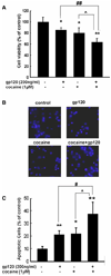
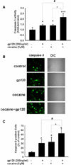
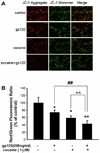
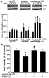
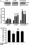
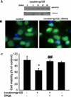
Although it has been well documented that drugs of abuse such as cocaine cause enhanced progression of human immunodeficiency virus (HIV)-associated neuropathological disorders, the underlying mechanisms mediating these effects remain poorly understood. The present study demonstrated that exposure of rat primary neurons to both cocaine and gp120 resulted in increased cell toxicity compared to cells treated with either factor alone. The combinatorial toxicity of cocaine and gp120 was accompanied by an increase in both caspase-3 activity and expression of the proapoptotic protein Bax. Furthermore, increased neurotoxicity in the presence of both the agents was associated with a concomitant increase in the production of intracellular reactive oxygen species and loss of mitochondrial membrane potential. Increased neurotoxicity mediated by cocaine and gp120 was ameliorated by NADPH oxidase inhibitor apocynin, thus underscoring the role of oxidative stress in this cooperation. Signaling pathways including c-jun N-teminal kinase (JNK), p38, extracellular signal-regulated kinase (ERK)/mitogen-activated protein kinases (MAPK), and nuclear factor (NF)-kappaB were also identified to be critical in the neurotoxicity induced by cocaine and gp120. These findings thus underscore the role of oxidative stress, mitochondrial and MAPK signal pathways in cocaine and HIV gp120-mediated neurotoxicity.
Figures




Similar articles
-
HIV-1 gp120 and drugs of abuse: interactions in the central nervous system.Curr HIV Res. 2012 Jul;10(5):369-83. doi: 10.2174/157016212802138724. Curr HIV Res. 2012. PMID: 22591361 Free PMC article. Review.
-
Cocaine potentiates astrocyte toxicity mediated by human immunodeficiency virus (HIV-1) protein gp120.PLoS One. 2010 Oct 15;5(10):e13427. doi: 10.1371/journal.pone.0013427. PLoS One. 2010. PMID: 20976166 Free PMC article.
-
Platelet-derived growth factor protects neurons against gp120-mediated toxicity.J Neurovirol. 2008 Jan;14(1):62-72. doi: 10.1080/13550280701809084. J Neurovirol. 2008. PMID: 18300076 Free PMC article.
-
Cocaine induces apoptosis in fetal rat myocardial cells through the p38 mitogen-activated protein kinase and mitochondrial/cytochrome c pathways.J Pharmacol Exp Ther. 2005 Jan;312(1):112-9. doi: 10.1124/jpet.104.073494. Epub 2004 Sep 13. J Pharmacol Exp Ther. 2005. PMID: 15365088
-
Cocaine and HIV-1 interplay in CNS: cellular and molecular mechanisms.Curr HIV Res. 2012 Jul;10(5):425-8. doi: 10.2174/157016212802138823. Curr HIV Res. 2012. PMID: 22591366 Free PMC article. Review.
Cited by
-
HIV-1 gp120 and drugs of abuse: interactions in the central nervous system.Curr HIV Res. 2012 Jul;10(5):369-83. doi: 10.2174/157016212802138724. Curr HIV Res. 2012. PMID: 22591361 Free PMC article. Review.
-
Cocaine-mediated induction of microglial activation involves the ER stress-TLR2 axis.J Neuroinflammation. 2016 Feb 9;13:33. doi: 10.1186/s12974-016-0501-2. J Neuroinflammation. 2016. PMID: 26860188 Free PMC article.
-
Cocaine hijacks σ1 receptor to initiate induction of activated leukocyte cell adhesion molecule: implication for increased monocyte adhesion and migration in the CNS.J Neurosci. 2011 Apr 20;31(16):5942-55. doi: 10.1523/JNEUROSCI.5618-10.2011. J Neurosci. 2011. PMID: 21508219 Free PMC article.
-
Cocaine promotes primary human astrocyte proliferation via JNK-dependent up-regulation of cyclin A2.Restor Neurol Neurosci. 2016 Nov 22;34(6):965-976. doi: 10.3233/RNN-160676. Restor Neurol Neurosci. 2016. PMID: 27834787 Free PMC article.
-
Protective effects of curcumin against human immunodeficiency virus 1 gp120 V3 loop-induced neuronal injury in rats.Neural Regen Res. 2012 Jan 25;7(3):171-5. doi: 10.3969/j.issn.1673-5374.2012.03.002. Neural Regen Res. 2012. PMID: 25767494 Free PMC article.
References
-
- Aksenov MY, Aksenova MV, Nath A, Ray PD, Mactutus CF, Booze RM. Cocaine-mediated enhancement of Tat toxicity in rat hippocampal cell cultures: the role of oxidative stress and D1 dopamine receptor. Neurotoxicology. 2006;27:217–228. - PubMed
-
- Ang E, Chen J, Zagouras P, Magna H, Holland J, Schaeffer E, Nestler EJ. Induction of nuclear factor-kappaB in nucleus accumbens by chronic cocaine administration. J Neurochem. 2001;79:221–224. - PubMed
-
- Anthony JC, Vlahov D, Nelson KE, Cohn S, Astemborski J, Solomon L. New evidence on intravenous cocaine use and the risk of infection with human immunodeficiency virus type 1. Am J Epidemiol. 1991;134:1175–1189. - PubMed
-
- Baldwin GC, Roth MD, Tashkin DP. Acute and chronic effects of cocaine on the immune system and the possible link to AIDS. J Neuroimmunol. 1998;83:133–138. - PubMed
-
- Bansal AK, Mactutus CF, Nath A, Maragos W, Hauser KF, Booze RM. Neurotoxicity of HIV-1 proteins gp120 and Tat in the rat striatum. Brain Res. 2000;879:42–49. - PubMed
Publication types
MeSH terms
Substances
Grants and funding
LinkOut - more resources
Full Text Sources
Medical
Research Materials
Miscellaneous

