. 2009 Apr;216(2):471-80.
doi: 10.1016/j.expneurol.2009.01.004.

Chronic spinal hemisection in rats induces a progressive decline in transmission in uninjured fibers to motoneurons

Affiliations

Chronic spinal hemisection in rats induces a progressive decline in transmission in uninjured fibers to motoneurons

Victor L Arvanian et al. Exp Neurol. 2009 Apr.

Abstract

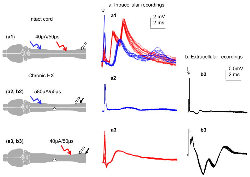
Although most spinal cord injuries are anatomically incomplete, only limited functional recovery has been observed in people and rats with partial lesions. To address why surviving fibers cannot mediate more complete recovery, we evaluated the physiological and anatomical status of spared fibers after unilateral hemisection (HX) of thoracic spinal cord in adult rats. We made intracellular and extracellular recordings at L5 (below HX) in response to electrical stimulation of contralateral white matter above (T6) and below (L1) HX. Responses from T6 displayed reduced amplitude, increased latency and elevated stimulus threshold in the fibers across from HX, beginning 1-2 weeks after HX. Ultrastructural analysis revealed demyelination of intact axons contralateral to the HX, with a time course similar to the conduction changes. Behavioral studies indicated partial recovery which arrested when conduction deficits began. In conclusion, this study is the first demonstration of the delayed decline of transmission through surviving axons to individual lumbar motoneurons during chronic stage of incomplete spinal cord injury in adult rats. These findings suggest a chronic pathological state in intact fibers and necessity for prompt treatment to minimize it.


Figures

Fig. 1
Fig. 1
Chronic injury, but not acute injury, induces decay of transmission to motoneurons through white matter contralateral to HX. Intracellular recordings were made in vivo from L5 motoneurons ipsilateral to T6 hemisection. Stimulation of ipsilateral and contralateral VLF at T6 above the level of injury. a–c: Responses from (a) ipsilateral and (b) contralateral VLF rostral to HX, and (c) ipsilateral VLF caudal to HX. Records displayed are among the largest responses for each group (average of 50 consecutive traces), with the arrow indicating the stimulus artifact at the left. Insets: superimposed 10 responses from same cell evoked by stimulation of the ipsilateral and contralateral VLF, respectively. Conditions: (i) intact spinal cord, (ii) same cord as above, but 10 minutes after HX, (iii) different cord, 4 wks post-HX. d: Position of the recording and stimulating electrodes. e: Reconstruction of the injury from T10 cross-sections from the cord presented in (iii); reconstruction from 3 sections 50 μM apart superimposed onto templates modified from (Paxinos et al., 1985). f: Summary of results demonstrating the decline in magnitude of motoneuron responses in L5 ipsilateral to HX at T10 (stimuli were delivered at T6 ipsilateral and contralateral to the HX, and at L1 ipsilateral to the HX; the symbols above some of the bars represent a significant decline from controls (p<0.05); all means were derived from n = 5–9 rats, and for each rat the response was an average of the maximum responses recorded intracellularly from 5 to 7 motoneurons).
Fig. 2
Fig. 2
Extracellular responses in L5 ventral horn contralateral to HX elicited by stimulation at T6 contralateral to HX. Diagrams show positions of the recording tungsten electrode (right-side ventral horn) and the stimulating electrode (right side VLF), stimulus intensities (at 1 Hz stimulation rate) and the lesion at T10. All superimposed traces are the successive responses evoked by stimuli of opposite polarity (50 consecutive traces each polarity, blue and red) We confirmed the neural basis of the recorded waves by demonstrating their invariance in response to changing stimulus polarity. a: Representative traces recorded in the intact cord. Note a marked fluctuation in both amplitude and latency in up-going waves (ii) and (iii), but not in the shortest-latency up-going wave (i), at a stimulus frequency of 1 Hz (these fluctuations of waves ii and iii were even more apparent when the stimulation frequency was raised to 10 Hz; not shown). b: Left hemisection (denoted as shaded triangle) did not induce marked changes of the evoked potentials conducted through the right uninjured side across from HX. c: Hemisection of left cord extended to include half the right cord (overhemisection) resulted in smaller amplitude responses. d: Complete transection of the cord (both left and right sides) completely abolished evoked responses, even at higher stimulus intensity.
Fig. 3
Fig. 3
Intracellular and extracellular recordings demonstrating conduction deficit and decay of transmission contralateral to chronic HX. Diagrams show position of recording electrodes (intracellular - clear arrow and extracellular - solid arrow) and stimulating electrodes (red and blue), and stimulus intensity required to evoke responses in uninjured and chronically injured spinal cords. a1: intact spinal cord. Stimulation of right VLF at T6 elicits EPSP in an L5 motoneuron on the same side that is similar in amplitude and of longer latency than the response elicited in the same motoneuron by stimulation at L1. Intracellular records showing the difference in EPSP latency were used for measurements of conduction velocity rather than extracellular records because the latter were often contaminated by the stimulus artifact particularly from the caudal position (L1). a2, b2: chronic left hemisected spinal cord (two weeks post-hemisection). Intracellular and extracellular responses at right L5 ventral horn elicited by stimulation of right VLF at T6 (a2, b2) are considerably smaller than the responses elicited at the same electrodes by stimulation at L1 (a3, b3).
Fig. 4
Fig. 4
Analysis of the total and myelinated axon number in the spared white matter on the intact side. A and B: Representative 60x (a) and Electron Microscope (b) images of the white matter (from labeled area in the insets). C: Number of myelinated axons in the spared white matter across from HX. Note marked decrease in the number of central myelinated axons in the spared white matter on the intact side in chronic (2 weeks and 6 weeks post-HX) vs acute HX rats (p < 0.05, one way non-parametric ANOVA).
Fig. 5
Fig. 5
Spontaneous recovery of locomotor function of both ipsilateral and contralateral to HX hindlimbs plateaus at about 2 weeks post-HX. In addition to BBB open-field locomotion score, more challenging tests, Narrowing Beam and Horizontal Irregular Ladder, were used to assess hindlimb function ipsilateral and contralateral to the lesion. Data plotted as means ± SEM with p<0.05 for all values at post-HX time points vs pre-operation values. BBB-scoring during 1st week post-HX revealed that all rats start with a very similar handicap (lesion severity). Note that function recovers spontaneously up to day 14 in all three tests, and after that the recovery plateaus.

References

    1. Antonino-Green DM, Cheng J, Magnuson DS. Neurons labeled from locomotor-related ventrolateral funiculus stimulus sites in the neonatal rat spinal cord. J Comp Neurol. 2002;442:226–38. - PubMed
    1. Arvanian VL, Bowers WJ, Anderson A, Horner PJ, Federoff HJ, Mendell LM. Combined delivery of neurotrophin-3 and NMDA receptors 2D subunit strengthens synaptic transmission in contused and staggered double hemisected spinal cord of neonatal rat. Exp Neurol. 2006;197:347–52. - PubMed
    1. Arvanian VL, Schnell L, Horner PJ, Bowers WJ, Federoff HJ, Schwab ME, Mendell LM. Combination Treatment With NT-3, NMDA-2d Subunits, And Anti-Nogo-A Antibody Increases Connections Of Transected Lateral Funiculus. Fibers To Lumbar Motoneurons Society For Neuroscience Abstracts #555.2 2006
    1. Ballermann M, Fouad K. Spontaneous locomotor recovery in spinal cord injured rats is accompanied by anatomical plasticity of reticulospinal fibers. Eur J Neurosci. 2006;23:1988–96. - PubMed
    1. Bareyre FM, Kerschensteiner M, Raineteau O, Mettenleiter TC, Weinmann O, Schwab ME. The injured spinal cord spontaneously forms a new intraspinal circuit in adult rats. Nat Neurosci. 2004;7:269–77. - PubMed

Publication types

MeSH terms