. 2009 Mar 24;7(3):e68.
doi: 10.1371/journal.pbio.1000068.

Quantifying the integration of quorum-sensing signals with single-cell resolution

Affiliations

Quantifying the integration of quorum-sensing signals with single-cell resolution

Tao Long et al. PLoS Biol. .

Abstract

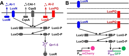
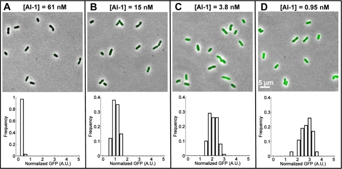
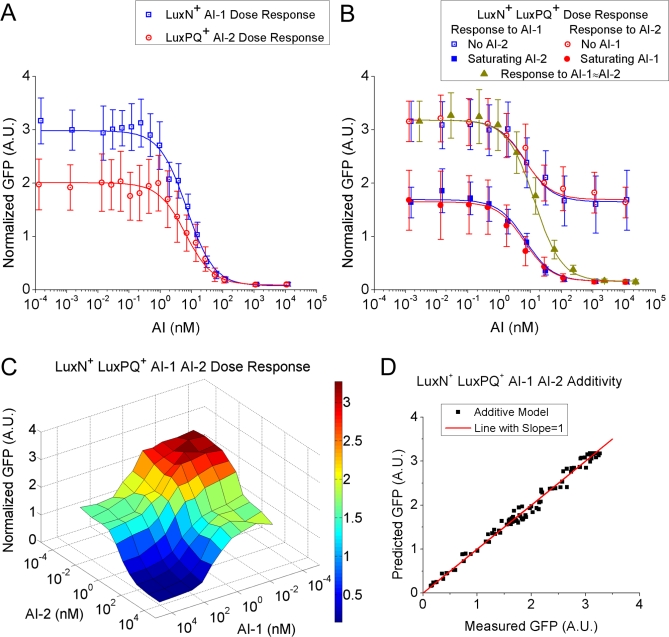
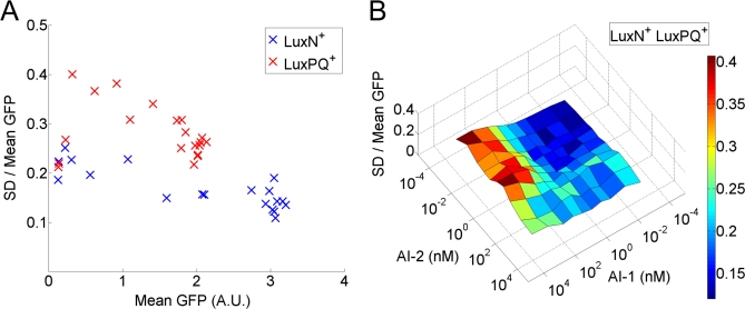
Cell-to-cell communication in bacteria is a process known as quorum sensing that relies on the production, detection, and response to the extracellular accumulation of signaling molecules called autoinducers. Often, bacteria use multiple autoinducers to obtain information about the vicinal cell density. However, how cells integrate and interpret the information contained within multiple autoinducers remains a mystery. Using single-cell fluorescence microscopy, we quantified the signaling responses to and analyzed the integration of multiple autoinducers by the model quorum-sensing bacterium Vibrio harveyi. Our results revealed that signals from two distinct autoinducers, AI-1 and AI-2, are combined strictly additively in a shared phosphorelay pathway, with each autoinducer contributing nearly equally to the total response. We found a coherent response across the population with little cell-to-cell variation, indicating that the entire population of cells can reliably distinguish several distinct conditions of external autoinducer concentration. We speculate that the use of multiple autoinducers allows a growing population of cells to synchronize gene expression during a series of distinct developmental stages.


Conflict of interest statement

Competing interests. The authors have declared that no competing interests exist.

Figures

Figure 1
Figure 1. The Quorum-Sensing Circuit of Wild-Type V. harveyi and Sensor Mutants Used in These Studies
(A) The wild-type quorum-sensing circuit consists of three parallel signaling pathways with three different autoinducers: AI-1, CAI-1, and AI-2. Their synthases are LuxM, CqsA, LuxS, and their transmembrane receptors are LuxN, CqsS, LuxPQ, respectively. In the absence of autoinducers (i.e., at low cell density), the receptors act predominantly as kinases and pass phosphate to LuxU and thence to LuxO. Phosphorylated-LuxO (LuxO-P) activates transcription of genes encoding five small regulatory RNAs (sRNAs). These sRNAs inhibit the translation of LuxR. In the presence of autoinducers (i.e., at high cell density), the receptors switch to a predominantly phosphatase-active state that reverses the direction of phosphoryl transfer through the circuit, so that LuxO is dephosphorylated and becomes inactive. Therefore, the genes encoding the five sRNAs are not transcribed, luxR mRNA is translated, and LuxR protein is made. (B) In the LuxN+ sensor mutant (top), the genes encoding cqsS, luxPQ, and the gene encoding the AI-1 synthase luxM are deleted. As a result, this mutant only responds to exogenously added AI-1. The LuxPQ+ sensor mutant (middle) responds exclusively to exogenous AI-2, and the LuxN+ LuxPQ+ sensor mutant (bottom) responds to exogenous AI-1 and AI-2. We quantify the responses using a qrr4-gfp transcriptional reporter fusion that is activated by LuxO-P. As an internal standard for fluorescence, the gene encoding mCherry is fused to a constitutive tac promoter and integrated at an intergenic region of the chromosome.
Figure 2
Figure 2. Single-Cell Microscopy Images and GFP Fluorescence Distributions of LuxN+ Cells of V. harveyi
Snapshots of cells growing exponentially at different AI-1 concentrations (indicated above images) and the corresponding GFP fluorescence distributions of single cells. The mean GFP fluorescence intensity of each cell is normalized by the cell's mean mCherry fluorescence intensity. Each distribution is obtained from 100 cells. A.U. denotes arbitrary units.
Figure 3
Figure 3. Autoinducer Dose Responses of V. harveyi Sensor Mutants
(A) Dose responses of LuxN+ cells to AI-1 (blue) and LuxPQ+ cells to AI-2 (red). Each average and standard deviation (error bar) of normalized GFP was obtained from microscopy images of 100 cells. Curves were fitted using αAI + βAI(1 + [AI]/K AI) with αAI-1 = 0.07, βAI-1 = 2.9, K AI-1 = 6.9 nM and αAI-2 = 0.09, βAI-2 = 1.9, K AI-2 = 6.4 nM. A.U. denotes arbitrary units. (B) Dose responses of LuxN+ LuxPQ+ cells to either AI-1 (blue) or AI-2 (red) while the other autoinducer is either absent (open squares and circles) or present at a saturating concentration (solid squares and circles). Data in yellow-green represent the response to approximately equal amounts of AI-1 and AI-2 (x-axis values indicate total autoinducer concentrations). (C) Dose–response surface of LuxN+ LuxPQ+ cells to various combinations of AI-1 and AI-2. Each vertex of the grid is the averaged normalized GFP fluorescence intensity obtained from a population of 100 cells exposed to the specified AI-1 and AI-2 concentrations. The dose–response curves in (B) correspond to cuts through this surface. (D) The response of LuxN+ LuxPQ+ cells to combined AI-1 and AI-2 shown in (C) can be well described by a simple additive model γ0 + γAI−1/(1 + [AI-1]/K AI-1) + γAI-2/(1 + [AI-2]/K AI-2), with γ0 = 0.16, γAI-1 = 1.53, γAI-2 = 1.49, K AI-1 = 6.9 nM, K AI-2 = 6.4 nM. The red line has a slope equal to one.
Figure 4
Figure 4. The Two Autoinducer-Sensing Pathways Contribute Differently to GFP-Expression Noise
(A) Relative noise, i.e., the standard deviation (SD) of the population divided by the mean, versus mean-normalized GFP fluorescence intensity for LuxN+ cells at different AI-1 concentrations (blue) and for LuxPQ+ cells at different AI-2 concentrations (red). (B) Relative noise for LuxN+ LuxPQ+ cells as a function of AI-1 and AI-2 concentrations.

Comment in

References

    1. Henke JM, Bassler BL. Bacterial social engagements. Trends Cell Biol. 2004;14:648–656. - PubMed
    1. Waters CM, Bassler BL. Quorum sensing: cell-to-cell communication in bacteria. Annu Rev Cell Dev Biol. 2005;21:319–346. - PubMed
    1. Bassler BL, Losick R. Bacterially speaking. Cell. 2006;125:237–246. - PubMed
    1. Chen X, Schauder S, Potier N, Van Dorsselaer A, Pelczer I, et al. Structural identification of a bacterial quorum-sensing signal-containing boron. Nature. 2002;415:545–549. - PubMed
    1. Xavier KB, Bassler BL. Interference with AI-2-mediated bacterial cell–cell communication. Nature. 2005;437:750–753. - PMC - PubMed

Publication types

MeSH terms