

Transforming growth factor-beta1 expression is up-regulated in maturation-stage enamel organ and may induce ameloblast apoptosis
- PMID: 19320718
- PMCID: PMC2711557
- DOI: 10.1111/j.1600-0722.2009.00612.x
Transforming growth factor-beta1 expression is up-regulated in maturation-stage enamel organ and may induce ameloblast apoptosis
Abstract
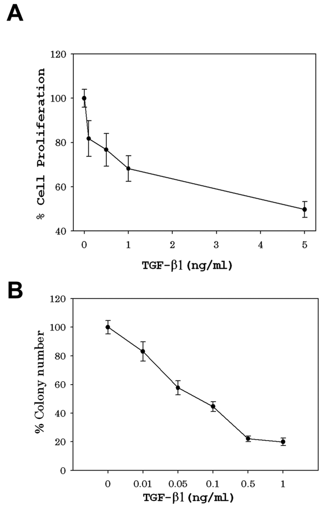
Transforming growth factor-beta1 (TGF-beta1) regulates a variety of cellular responses that are dependent on the developmental stage and on the origins of the cell or the tissue. In mature tissues, and especially in tissues of epithelial origin, TGF-beta1 is generally considered to be a growth inhibitor that may also promote apoptosis. The ameloblast cells of the enamel organ epithelium are adjacent to and responsible for the developing enamel layer on unerupted teeth. Once the enamel layer reaches its full thickness, the tall columnar secretory-stage ameloblasts shorten, and a portion of these maturation-stage ameloblasts become apoptotic. Here we investigate whether TGF-beta1 plays a role in apoptosis of the maturation-stage ameloblasts. We demonstrate in vitro that ameloblast lineage cells are highly susceptible to TGF-beta1-mediated growth arrest and are prone to TGF-beta1-mediated cell death/apoptosis. We also demonstrate in vivo that TGF-beta1 is expressed in the maturation-stage enamel organ at significantly higher levels than in the earlier secretory-stage enamel organ. This increased expression of TGF-beta1 correlates with an increase in expression of the enamel organ immediate-early stress-response gene and with a decrease in the anti-apoptotic Bcl2 : Bax expression ratio. We conclude that TGF-beta1 may play an important role in ameloblast apoptosis during the maturation stage of enamel development.
Figures


Similar articles
-
E-cadherin can replace N-cadherin during secretory-stage enamel development.PLoS One. 2014 Jul 11;9(7):e102153. doi: 10.1371/journal.pone.0102153. eCollection 2014. PLoS One. 2014. PMID: 25014356 Free PMC article.
-
The immunohistochemical localization of Bax and Bcl-2 and their relation to apoptosis during amelogenesis in developing rat molars.Arch Oral Biol. 2001 Jun;46(6):557-68. doi: 10.1016/s0003-9969(00)00139-4. Arch Oral Biol. 2001. PMID: 11311203
-
RUNX2 contributes to TGF-β1-induced expression of Wdr72 in ameloblasts during enamel mineralization.Biomed Pharmacother. 2019 Oct;118:109235. doi: 10.1016/j.biopha.2019.109235. Epub 2019 Jul 21. Biomed Pharmacother. 2019. PMID: 31336344
-
V-type ATPase proton pump expression during enamel formation.Matrix Biol. 2016 May-Jul;52-54:234-245. doi: 10.1016/j.matbio.2015.11.004. Epub 2015 Nov 14. Matrix Biol. 2016. PMID: 26586472 Free PMC article. Review.
-
Cellular and chemical events during enamel maturation.Crit Rev Oral Biol Med. 1998;9(2):128-61. doi: 10.1177/10454411980090020101. Crit Rev Oral Biol Med. 1998. PMID: 9603233 Review.
Cited by
-
Transcription factor FoxO1 is essential for enamel biomineralization.PLoS One. 2012;7(1):e30357. doi: 10.1371/journal.pone.0030357. Epub 2012 Jan 24. PLoS One. 2012. PMID: 22291941 Free PMC article.
-
TGF-β1/Smad3 Signaling Is Required to Alleviate Fluoride-Induced Enamel Hypomineralization.Biol Trace Elem Res. 2024 Feb;202(2):569-579. doi: 10.1007/s12011-023-03688-y. Epub 2023 May 4. Biol Trace Elem Res. 2024. PMID: 37140770
-
The role of the TGF-β1 signaling pathway in the process of amelogenesis.Front Physiol. 2025 Apr 9;16:1586769. doi: 10.3389/fphys.2025.1586769. eCollection 2025. Front Physiol. 2025. PMID: 40271211 Free PMC article. Review.
-
Multiple Calcium Export Exchangers and Pumps Are a Prominent Feature of Enamel Organ Cells.Front Physiol. 2017 May 23;8:336. doi: 10.3389/fphys.2017.00336. eCollection 2017. Front Physiol. 2017. PMID: 28588505 Free PMC article.
-
Apoptotic signaling in mouse odontogenesis.OMICS. 2012 Jan-Feb;16(1-2):60-70. doi: 10.1089/omi.2011.0039. Epub 2011 Dec 28. OMICS. 2012. PMID: 22204278 Free PMC article. Review.
References
-
- Cole AS, Eastoe J. Biochemistry and Oral Biology. London: Butterworth & Co. LTD; 1988.
-
- Bartlett JD, Simmer JP. Proteinases in Developing Dental Enamel. Crit Rev Oral Biol Med. 1999;10:425–441. - PubMed
-
- Smith CE. Cellular and Chemical Events During Enamel Maturation. Crit Rev Oral Biol Med. 1998;9:128–161. - PubMed
-
- Kallenbach E. Fine Structure of Rat Incisor Enamel Organ During Late Pigmentation and Regression Stages. J Ultrastruct Res. 1970;30:38–63. - PubMed
-
- Smith CE, Warshawsky H. Quantitative Analysis of Cell Turnover in the Enamel Organ of the Rat Incisor. Evidence for Ameloblast Death Immediately After Enamel Matrix Secretion. Anat Rec. 1977;187:63–98. - PubMed
Publication types
MeSH terms
Substances
Grants and funding
LinkOut - more resources
Full Text Sources
Research Materials
