

Gpnmb is a melanosome-associated glycoprotein that contributes to melanocyte/keratinocyte adhesion in a RGD-dependent fashion
- PMID: 19320736
- PMCID: PMC2774115
- DOI: 10.1111/j.1600-0625.2008.00830.x
Gpnmb is a melanosome-associated glycoprotein that contributes to melanocyte/keratinocyte adhesion in a RGD-dependent fashion
Abstract
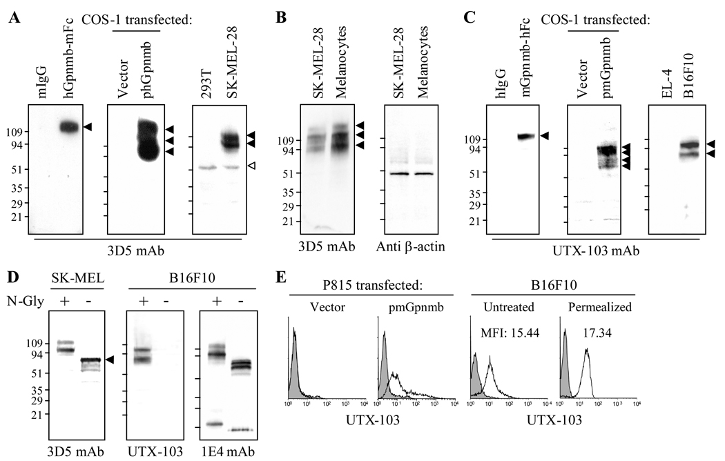
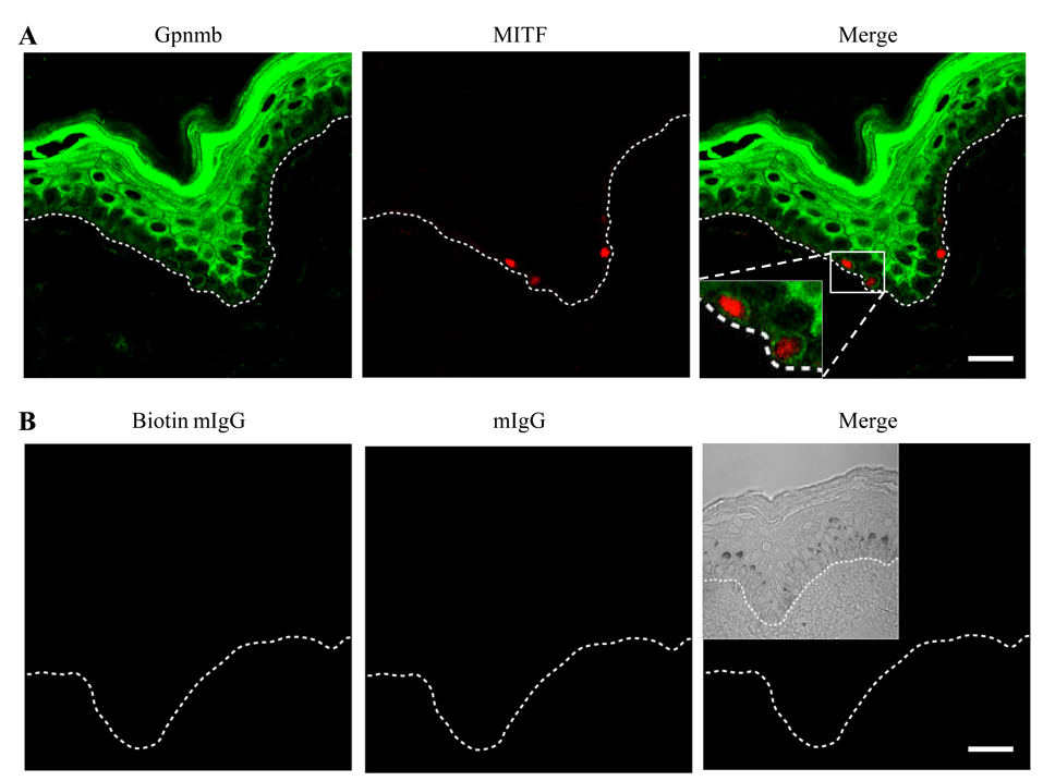
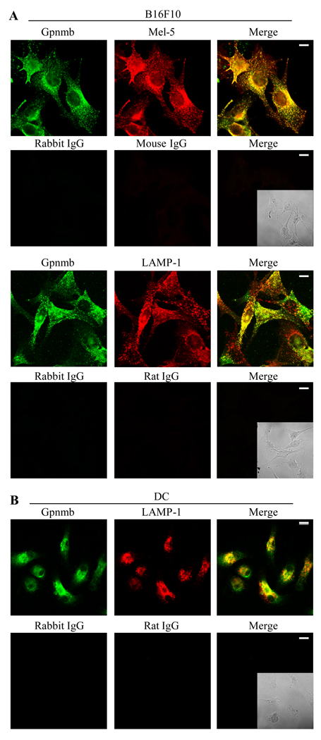
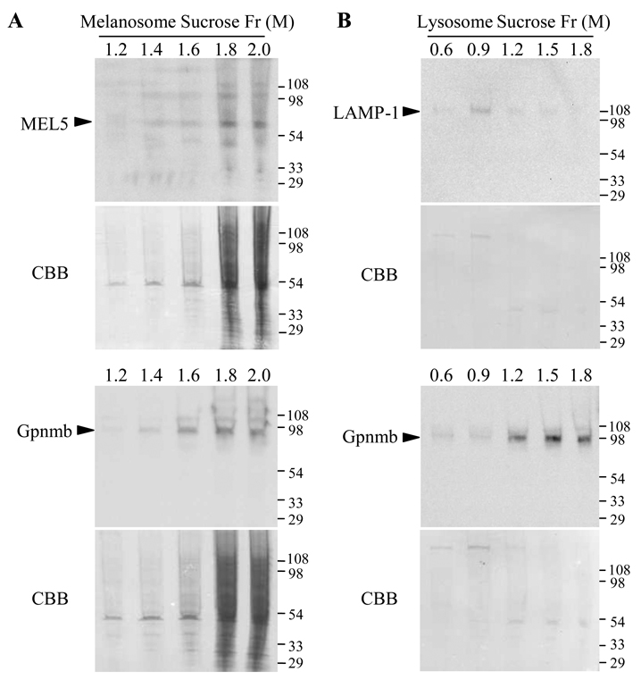
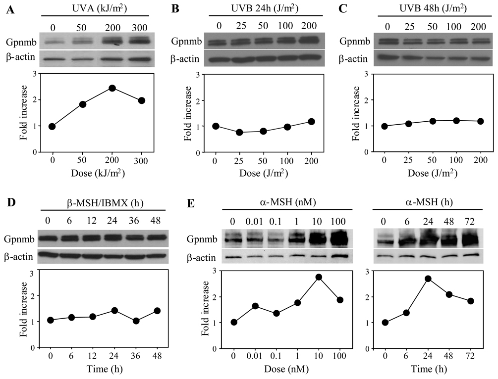
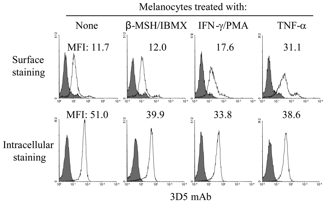
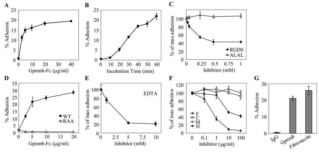
Gpnmb is a glycosylated transmembrane protein implicated in the development of glaucoma in mice and melanoma in humans. It shares significant amino acid sequence homology with the melanosome protein Pmel-17. Its extracellular domain contains a RGD motif for binding to integrin and its intracellular domain has a putative endosomal and/or melanosomal-sorting motif. These features led us to posit that Gpnmb is associated with melanosomes and involved in cell adhesion. We showed that human Gpnmb is expressed constitutively by melanoma cell lines, primary-cultured melanocytes and epidermal melanocytes in situ, with most of it found intracellularly within melanosomes and to a lesser degree in lysosomes. Our newly developed monoclonal antibody revealed surface expression of Gpnmb on these pigment cells, albeit to a lesser degree than the intracellular fraction. Gpnmb expression was upregulated by UVA (but not UVB) irradiation and by alpha-melanocyte-stimulating hormone (MSH) (but not beta-MSH); its cell surface expression on melanocytes (but not on melanoma cells) was increased markedly by IFN-gamma and TNF-alpha. PAM212 keratinocytes adhered to immobilized Gpnmb in a RGD-dependent manner. These results indicate that Gpnmb is a melanosome-associated glycoprotein that contributes to the adhesion of melanocytes with keratinocytes.
Figures
Similar articles
-
Glycoprotein nonmetastatic melanoma protein b, a melanocytic cell marker, is a melanosome-specific and proteolytically released protein.FASEB J. 2010 May;24(5):1616-29. doi: 10.1096/fj.09-151019. Epub 2010 Jan 7. FASEB J. 2010. PMID: 20056711 Free PMC article.
-
Silencing of GPNMB by siRNA inhibits the formation of melanosomes in melanocytes in a MITF-independent fashion.PLoS One. 2012;7(8):e42955. doi: 10.1371/journal.pone.0042955. Epub 2012 Aug 13. PLoS One. 2012. PMID: 22912767 Free PMC article.
-
A method for quantifying melanosome transfer efficacy from melanocytes to keratinocytes in vitro.Pigment Cell Melanoma Res. 2008 Oct;21(5):559-64. doi: 10.1111/j.1755-148X.2008.00487.x. Epub 2007 Jul 9. Pigment Cell Melanoma Res. 2008. PMID: 18702635
-
Keratinocyte-melanocyte interactions during melanosome transfer.Pigment Cell Res. 2001 Aug;14(4):236-42. doi: 10.1034/j.1600-0749.2001.140402.x. Pigment Cell Res. 2001. PMID: 11549105 Review.
-
Adhesion, migration and communication in melanocytes and melanoma.Pigment Cell Res. 2005 Jun;18(3):150-9. doi: 10.1111/j.1600-0749.2005.00235.x. Pigment Cell Res. 2005. PMID: 15892711 Review.
Cited by
-
The PKD domain distinguishes the trafficking and amyloidogenic properties of the pigment cell protein PMEL and its homologue GPNMB.Pigment Cell Melanoma Res. 2013 Jul;26(4):470-86. doi: 10.1111/pcmr.12084. Epub 2013 Apr 2. Pigment Cell Melanoma Res. 2013. PMID: 23452376 Free PMC article.
-
The DC-HIL/syndecan-4 pathway regulates autoimmune responses through myeloid-derived suppressor cells.J Immunol. 2014 Mar 15;192(6):2576-84. doi: 10.4049/jimmunol.1301857. Epub 2014 Feb 10. J Immunol. 2014. PMID: 24516197 Free PMC article.
-
GPNMB is expressed in human epidermal keratinocytes but disappears in the vitiligo lesional skin.Sci Rep. 2020 Mar 18;10(1):4930. doi: 10.1038/s41598-020-61931-1. Sci Rep. 2020. PMID: 32188902 Free PMC article.
-
Loss of GPNMB Causes Autosomal-Recessive Amyloidosis Cutis Dyschromica in Humans.Am J Hum Genet. 2018 Feb 1;102(2):219-232. doi: 10.1016/j.ajhg.2017.12.012. Epub 2018 Jan 11. Am J Hum Genet. 2018. PMID: 29336782 Free PMC article.
-
Functional Domains and Evolutionary History of the PMEL and GPNMB Family Proteins.Molecules. 2021 Jun 9;26(12):3529. doi: 10.3390/molecules26123529. Molecules. 2021. PMID: 34207849 Free PMC article.
References
-
- Shikano S, Bonkobara M, Zukas PK, Ariizumi K. Molecular cloning of a dendritic cell-associated transmembrane protein, DC-HIL, that promotes RGD-dependent adhesion of endothelial cells through recognition of heparan sulfate proteoglycans. J Biol Chem. 2001;276:8125–8134. - PubMed
-
- Weterman MA, Ajubi N, van Dinter I, et al. nmb, a novel gene, is expressed in low-metastatic human melanoma cell lines and xenografts. Int J Cancer. 1995;60:73–81. - PubMed
-
- Anderson MG, Smith RS, Hawes NL, et al. Mutations in genes encoding melanosomal proteins cause pigmentary glaucoma in DBA/2J mice. Nat Genet. 2002;30:81–85. - PubMed
-
- Safadi FF, Xu J, Smock SL, et al. Cloning and characterization of osteoactivin, a novel cDNA expressed in osteoblasts. J Cell Biol. 2001;84:12–26. - PubMed
Publication types
MeSH terms
Substances
Grants and funding
LinkOut - more resources
Full Text Sources
Other Literature Sources
Medical
