

Cellular/intramuscular myxoma and grade I myxofibrosarcoma are characterized by distinct genetic alterations and specific composition of their extracellular matrix
- PMID: 19320777
- PMCID: PMC4496143
- DOI: 10.1111/j.1582-4934.2009.00747.x
Cellular/intramuscular myxoma and grade I myxofibrosarcoma are characterized by distinct genetic alterations and specific composition of their extracellular matrix
Abstract
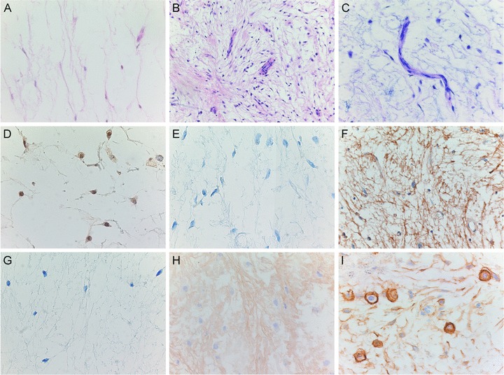
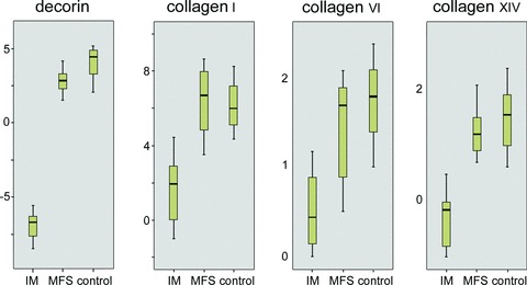
Cellular myxoma and grade I myxofibrosarcoma are mesenchymal tumours that are characterized by their abundant myxoid extracellular matrix (ECM). Despite their histological overlap, they differ clinically. Diagnosis is therefore difficult though important. We investigated their (cyto) genetics and ECM. GNAS1-activating mutations have been described in intramuscular myxoma, and lead to downstream activation of cFos. KRAS and TP53 mutations are commonly involved in sarcomagenesis whereby KRAS subsequently activates c-Fos. A well-documented series of intramuscular myxoma (three typical cases and seven cases of the more challenging cellular variant) and grade I myxofibrosarcoma (n = 10) cases were karyotyped, analyzed for GNAS1, KRAS and TP53 mutations and downstream activation of c-Fos mRNA and protein expression. ECM was studied by liquid chromatography mass spectrometry and expression of proteins identified was validated by immunohistochemistry and qPCR. Grade I myxofibrosarcoma showed variable, non-specific cyto-genetic aberrations in 83,5% of cases (n = 6) whereas karyotypes of intramuscular myxoma were all normal (n = 7). GNAS1-activating mutations were exclusively found in 50% of intramuscular myxoma. Both tumour types showed over-expression of c-Fos mRNA and protein. No mutations in KRAS codon 12/13 or in TP53 were detected. Liquid chromatography mass spectrometry revealed structural proteins (collagen types I, VI, XII, XIV and decorin) in grade I myxofibrosarcoma lacking in intramuscular myxoma. This was confirmed by immunohistochemistry and qPCR. Intramuscular/cellular myxoma and grade I myxofibrosarcoma show different molecular genetic aberrations and different composition of their ECM that probably contribute to their diverse clinical behaviour. GNAS1 mutation analysis can be helpful to distinguish intramuscular myxoma from grade I myxofibrosarcoma in selected cases.
Figures
References
-
- Graadt van Roggen JF, Hogendoorn PCW, Fletcher CDM. Myxoid tumours of soft tissue. Histopathology. 1999;35:291–12. - PubMed
-
- Graadt van Roggen JF, McMenamin ME, Fletcher CDM. Cellular myxoma: a clinicopathologic study of 38 cases confirming indolent clinical behavior. Mod Pathol. 2001;39:287–97. - PubMed
-
- Mentzel T, Calonje E, Wadden C, et al. Myxofibrosarcoma. Clinicopathologic analysis of 75 cases with emphasis on the low-grade variant. Am J Surg Pathol. 1996;20:391–05. - PubMed
-
- Meis-Kindblom JM, Sjogren H, Kindblom LG, et al. Cytogenetic and molecular genetic analyses of liposarcoma and its soft tissue simulators: recognition of new variants and differential diagnosis. Virchows Arch. 2001;439:141–51. - PubMed
-
- Willems SM, Debiec-Rychter M, Szuhai K, et al. Local recurrence of myxofibrosarcoma is associated with increase in tumour grade and cytogenetic aberrations, suggesting a multistep tumour progression model. Mod Pathol. 2006;19:407–16. - PubMed
Publication types
MeSH terms
Substances
LinkOut - more resources
Full Text Sources
Research Materials
Miscellaneous
