. 2009 Apr;72(2):491-505.
doi: 10.1111/j.1365-2958.2009.06664.x. Epub 2009 Mar 17.

HlyU acts as an H-NS antirepressor in the regulation of the RTX toxin gene essential for the virulence of the human pathogen Vibrio vulnificus CMCP6

Affiliations

HlyU acts as an H-NS antirepressor in the regulation of the RTX toxin gene essential for the virulence of the human pathogen Vibrio vulnificus CMCP6

Moqing Liu et al. Mol Microbiol. 2009 Apr.

Abstract

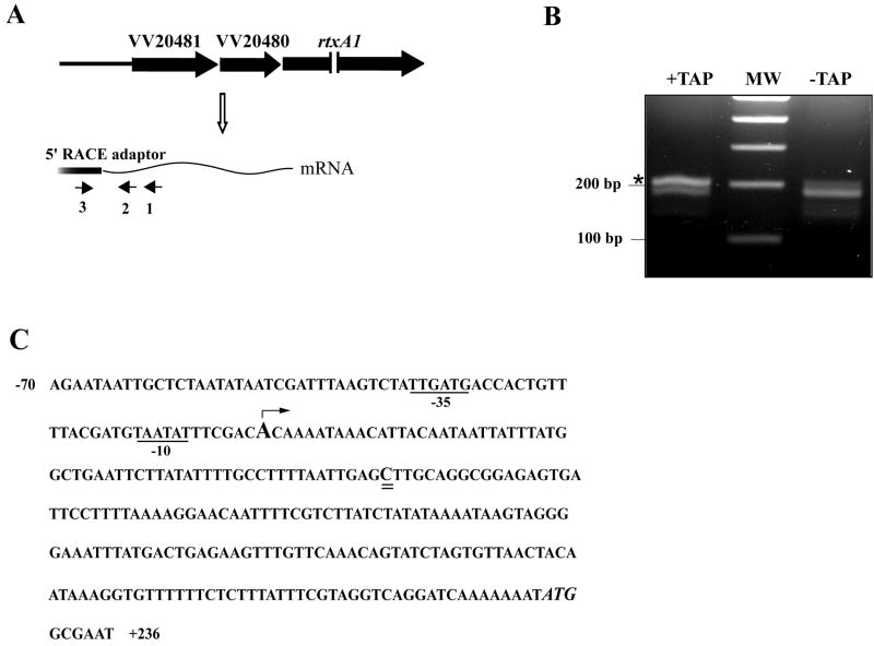
In Vibrio vulnificus, HlyU upregulates the expression of the large RTX toxin gene. In this work we identified the binding site of HlyU to -417 to -376 bp of the rtxA1 operon transcription start site. lacZ fusions for a series of progressive deletions from the rtxA1 operon promoter showed that transcriptional activity increased independently of HlyU when its binding site was absent. Thus HlyU must regulate the rtxA1 operon expression by antagonizing a negative regulator. Concomitantly we found that an hns mutant resulted in an increase in the expression of the rtxA1 operon genes. Multiple copies of HlyU can increase the promoter activity only in the presence of H-NS underscoring the hypothesis that HlyU must alleviate the repression by this protein. H-NS binds to a region that extends upstream and downstream of the rtxA1 operon promoter. In the upstream region it binds to five AT-rich sites of which two overlap the HlyU binding site. Competitive footprinting and gel shift data demonstrate HlyU's higher affinity as compared with H-NS resulting in the de-repression and a corresponding increased expression of the rtxA1 operon.


Figures

Fig.1
Fig.1
The modified RLM-RACE to determine the transcription start site of the rtxA1 operon. (A) Location of the primers in the RLM-RACE experiment. Small arrows represent the location of primers. Primer 1, P20481-R; primer 2, RACE20481-R-in; primer 3, 5’RACE outer primer. (B) The PCR results from primer 3 (the adapter-specific 5’ RACE outer primer) and primer 2; the asterisk points out the extra band from the TAP-treated sample compared with the TAP-untreated sample. MW, DNA molecular weight marker. (C) Sequence analysis of the upstream region of the rtxA1 operon promoter. The transcription start site is indicated by the bent arrow. The -10 and -35 regions are underlined. The processing point of the mRNA is double underlined, and the putative translation start site is in italics.
Fig. 2
Fig. 2
Ribonuclease protection assay (RPA) to detect other transcript(s) initiated between the identified transcription start site and the HlyU binding site. (A) Location of the probes used in RPA: probe a’, +91 to -69; probe b’, -99 to −278, and probe c’, -359 to -523. The capital “A” stands for the transcription start site; the black bar represents the HlyU binding site. (B) The results from the RPA. The total RNA from the wild type CMCP6 cells was hybridized with probes a’, b’ and c’ respectively, then treated with RNase A and RNase T1. Lanes 1, 3 and 5 are the ribonuclease protection results for probe a’, b’ and c’ respectively; lanes 2, 4 and 6 stand for a’, b’ and c’ probe only, respectively.
Fig. 3
Fig. 3
DNase I protection by HlyU at the upstream region of the rtxA1 operon promoter. (A) Location of the DNA probe fragments a (+279 to +89), b (+111 to -29), c (-30 to -279), and d (-260 to -523). (B) DNase I footpringting assay by HlyU and fragment d. The coding strand of the fragment extending from -523 to -260 bp of the transcription start site was incubated with increasing concentrations of HlyU and treated with DNase I. Lane 1, no protein; lane 2-3, HlyU, 25 nM and 100 nM, respectively. Lanes A, C, G and T represent the nucleotide sequencing ladders of pTopo-FP-P20481. The open bar indicated the protection region from 417 to 376 bp upstream of the transcription start site with the sequence TGTAATTATTAGTTTTTGTTAAATTAGCATTTTCTTTAAATT.
Fig. 4
Fig. 4
Promoter activities of various lacZ transcriptional fusions in the hlyU+lacZ) and hns-lacZ ΔhlyU) strains. The figures described the promoter fragments which were fused to lacZ gene in pTL-523, pTL-428, pTL-347, pTL-278 and pTL-35, repectively (from up to down). The numbers in the right panel represents β-galactosidase activities for these fusion plasmids in the strains indicated. β-galactosidase activities were determined as described by Miller (Miller, 1992). The data are mean values from 3 independent experiments (± standard deviation). ΔlacZ, ALE-LAC; ΔlacZ ΔhlyU, MQ4.
Fig. 5
Fig. 5
β-galactosidase activity assay of the V. vulnificus hns+lacZ) and hns-lacZ hns∷pDM4) strains harboring the lacZ-fusion plasmid and the hlyU multicopy plasmid. ΔlacZ, ALE-LAC; ΔlacZ hns∷pDM4, MQ5; pTL-523, lacZ fusion plasmid; pMMB, pMMB208K; pMMB-hlyU, pMMB208K derivative containing the wild-type hlyU gene. β-galactosidase activities were determined as described by Miller (Miller, 1992). The data are mean values from 3 independent experiments (± standard deviation).
Fig. 6
Fig. 6
H-NS binds to the far upstream region of the rtxA1 operon promoter. (A) Gel mobility shift assay for the binding of the H-NS protein to the fragment d. The labeled DNA fragment (5 nM) was mixed with varying concentrations of H-NS protein as indicated. For the competition analysis, the unlabeled DNA was used as a competitor. Before addition of 1 μM H-NS, 300 nM of unlabeled specific competitor DNA (lane 12) or 300 nM of unlabeled non-specific competitor DNA (lane 13) which is from 150 bp of rtxA1 gene was added to the reaction containing a 5 nM concentration of the labeled DNA probe. (B) Identification of H-NS binding sites using DNase I footprint analysis. The coding strand of DNA relative to the fragment d was incubated with increasing concentrations of H-NS and treated with DNase I. Lane 1, no protein; lane 2-6, H-NS (0.2, 0.5, 0.8, 1.5, 3.0 μM respectively). Lanes A, C, G and T represent the nucleotide sequencing ladders of pTopo-FP-P20481. The black bars (I, II, III, IV and V) indicate the H-NS protection sites. (C) The sequence of H-NS protection region in the DNase I footprinting analysis is underlined and the region protected by HlyU is in italics.
Fig. 7
Fig. 7
H-NS binds to the region from upstream to downstream of the rtxA1 operon promoter. Gel mobility shift assay for the binding of the H-NS protein to the fragment a, b, and c. The labeled DNA fragment (5 nM) was mixed with varying concentrations of H-NS protein as indicated. For the competition analysis, the excess of unlabeled specific DNA was added to the reaction before the addition of 1 μM H-NS (lane 4).
Fig. 8
Fig. 8
Competitive mobility shift assay between HlyU and H-NS on the upstream region of the rtxA1 operon promoter. (A) Increasing concentrations of HlyU were added to a fixed concentration of H-NS and DNA (fragment d). (B) Increasing concentrations of H-NS were added to a fixed concentration of HlyU and DNA (fragment d).
Fig. 9
Fig. 9
Competitive DNase I footprinting assays between HlyU and H-NS on the upstream region of the rtxA1 operon promoter. (A) Increasing concentrations of HlyU and the fixed concentration of H-NS. Lane 1 and 2, no protein; lane 3, HlyU only (0.2 μM); lane 4, H-NS only (1.0 μM); lane 5-8, HlyU (0.2, 0.8, 1.5, 3.0 μM repectively) with the fixed concentration of H-NS (1.0 μM). (B) Increasing concentrations of H-NS and the fixed concentration of HlyU. Lane 1 and 2, no protein; lane 3, H-NS only (1.0 μM); lane 4, HlyU only (0.2 μM); lane 5-8, H-NS (0.2, 0.8, 1.5 and 3.0 μM repectively) with the fixed concentration of HlyU (0.2 μM). Lanes A, C, G and T represent the nucleotide sequencing ladders of pTopo-FP-P20481. Regions protected by H-NS are shown by the black bar, and region protected by HlyU is shown by the open bar.

Similar articles

Cited by

References

    1. Alice AF, Naka H, Crosa JH. Global gene expression as a function of the iron status of the bacterial cell: influence of differentially expressed genes in the virulence of the human pathogen Vibrio vulnificus. Infect Immun. 2008;76:4019–4037. - PMC - PubMed
    1. Amaro C, Biosca EG, Fouz B, Alcaide E, Esteve C. Evidence that water transmits Vibrio vulnificus biotype 2 infections to eels. Appl Environ Microbiol. 1995;61:1133–1137. - PMC - PubMed
    1. Banos RC, Pons JI, Madrid C, Juarez A. A global modulatory role for the Yersinia enterocolitica H-NS protein. Microbiology. 2008;154:1281–1289. - PubMed
    1. Beloin C, Dorman CJ. An extended role for the nucleoid structuring protein H-NS in the virulence gene regulatory cascade of Shigella flexneri. Mol Microbiol. 2003;47:825–838. - PubMed
    1. Bensing BA, Meyer BJ, Dunny GM. Sensitive detection of bacterial transcription initiation sites and differentiation from RNA processing sites in the pheromone-induced plasmid transfer system of Enterococcus faecalis. Proc Natl Acad Sci U S A. 1996;93:7794–7799. - PMC - PubMed

Publication types

MeSH terms

LinkOut - more resources