. 2009 Mar 25:5:14.
doi: 10.1186/1744-8069-5-14.

Increased peripheral nerve excitability and local NaV1.8 mRNA up-regulation in painful neuropathy

Affiliations

Increased peripheral nerve excitability and local NaV1.8 mRNA up-regulation in painful neuropathy

Devang Kashyap Thakor et al. Mol Pain. .

Abstract

Background: Neuropathic pain caused by peripheral nerve injury is a chronic disorder that represents a significant clinical challenge because the pathological mechanisms have not been fully elucidated. Several studies have suggested the involvement of various sodium channels, including tetrodotoxin-resistant NaV1.8, in affected dorsal root ganglion (DRG) neurons. We have hypothesized that altered local expression of NaV1.8 in the peripheral axons of DRG neurons could facilitate nociceptive signal generation and propagation after neuropathic injury.

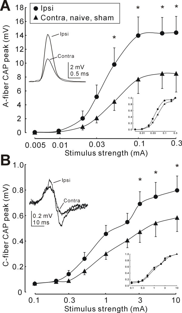
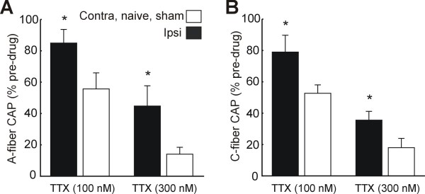
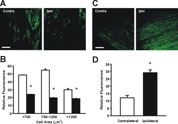
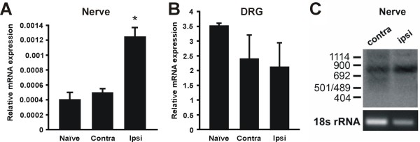
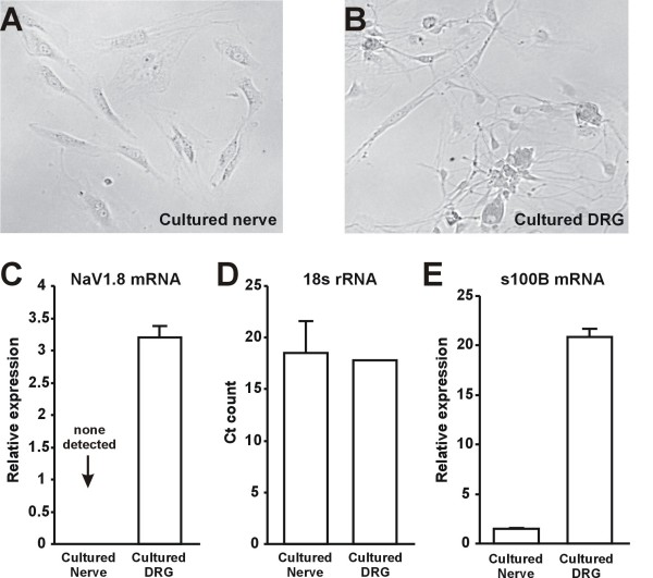
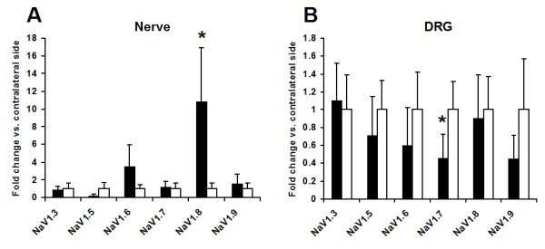
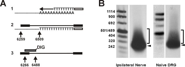
Results: After unilateral sciatic nerve entrapment injury in rats, compound action potential amplitudes were increased in both myelinated and unmyelinated fibers of the ipsilateral sciatic nerve. Tetrodotoxin resistance of both fiber populations and sciatic nerve NaV1.8 immunoreactivity were also increased. Further analysis of NaV1.8 distribution revealed that immunoreactivity and mRNA levels were decreased and unaffected, respectively, in the ipsilateral L4 and L5 DRG; however sciatic nerve NaV1.8 mRNA showed nearly an 11-fold ipsilateral increase. Nav1.8 mRNA observed in the sciatic nerve was likely of axonal origin since it was not detected in non-neuronal cells cultured from nerve tissue. Absence of changes in NaV1.8 mRNA polyadenylation suggests that increased mRNA stability was not responsible for the selective peripheral mRNA increase. Furthermore, mRNA levels of NaV1.3, NaV1.5, NaV1.6, NaV1.7, and NaV1.9 were not significantly different between ipsilateral and contralateral nerves. We therefore propose that selective NaV1.8 mRNA axonal transport and local up-regulation could contribute to the hyperexcitability of peripheral nerves in some neuropathic pain states.

Conclusion: Cuff entrapment injury resulted in significantly elevated axonal excitability and increased NaV1.8 immunoreactivity in rat sciatic nerves. The concomitant axonal accumulation of NaV1.8 mRNA may play a role in the pathogenesis of this model of neuropathic pain.


Figures

Figure 1
Figure 1
Increased excitability of ipsilateral sciatic nerve after SNE. A: Stimulus-output plot of A-fiber CAP peak for sciatic nerve ipsilateral to SNE (n = 8) compared to preparations from contralateral, naïve or sham control rats (n = 8). Inset (left) shows examples of ipsilateral and contralateral A-fiber CAP with peaks aligned for clarity (0.1 mA stimulus). *, p < 0.05 (two-way ANOVA, Tukey post-hoc). Inset (bottom right) shows normalized data with error bars removed for clarity. Note the leftward shift of data from ipsilateral nerves. B: Stimulus-output plot of C-fiber CAP peak for sciatic nerve ipsilateral to SNE compared to preparations from contralateral, naïve or control rats. Inset (left) shows examples of ipsilateral and contralateral C-fiber CAP (10 mA stimulus). Inset (bottom right) shows normalized data with error bars removed for clarity. Stimulus durations were 0.1 ms and 0.5 ms for A- and C-fiber CAP, respectively throughout. Tissue was harvested 2 weeks after SNE or sham surgery.
Figure 2
Figure 2
Increased TTX-resistance of the sciatic nerve after SNE. A: TTX-resistance of the A-fiber sciatic nerve compound action potential (CAP) ipsilateral to SNE (n = 8) compared to contralateral, naïve and sham surgery nerves (n = 8). CAP response is expressed as a % of recorded amplitude before TTX administration. A-fiber CAP stimulus was 0.1 mA, 0.1 ms throughout. B: Changes in TTX-resistance of the C-fiber sciatic nerve CAP after SNE. C-fiber CAP stimulus was 10 mA, 0.5 ms throughout. * indicates significant difference from control nerve sensitivity to TTX (p < 0.05, two-way ANOVA, Tukey post-hoc). Tissue was harvested 2 weeks after SNE or sham surgery.
Figure 3
Figure 3
NaV1.8 protein is down-regulated in the DRG and up-regulated in the nerve after SNE. A: Nav1.8-ir in representative DRG sections after SNE. B: Quantification of fluorescence intensity in DRG neurons. Neurons are classified by cell area. □: contralateral. ▪: ipsilateral. DRG measurements were from 3 contralateral and 6 ipsilateral sections (n = 3. *; p < 0.05, one-way ANOVA, Tukey post-hoc). C: NaV1.8-ir in representative sections of sciatic nerve after SNE. D: Quantification of fluorescence intensity in sciatic nerve. Measurements were made from 8 contralateral and 8 ipsilateral sections. (n = 3, *; p < 0.05, t-test). Scale bar: 100 μm for A and C. Tissue was harvested 2 weeks after SNE. For both DRG and nerve sections, average intensity was measured using ImageJ software. Output intensity values were on a 0–255 scale based on the 255 shades of grey present in the image, with 0 representing black and 255 representing white. The output numerical intensity value was defined as "relative fluorescence."
Figure 4
Figure 4
Detection of NaV1.8 mRNA in the sciatic nerve. A. NaV1.8 mRNA levels were significantly increased in the sciatic nerve ipsilateral to the injury site as compared to contralateral and uninjured DRG. (n = 3; p < 0.05, one-way ANOVA with Tukey post-hoc). B. NaV1.8 mRNA levels in SNE-injured DRG were not significantly different from those of uninjured DRG (n = 4 for uninjured DRG and n = 3 for all other samples). C: Southern blot of the Nav1.8 3' RACE product from contralateral and ipsilateral sciatic nerve. Note the presence of a distinct band representing the expected 3'RACE product of 830 bp. Sequencing of the RACE product confirmed the presence of the 3' end of the NaV1.8 coding region as well as the NaV1.8 3' un-translated region and polyA tail. Tissue was harvested 2 weeks after SNE.
Figure 5
Figure 5
Axonal origin of sciatic nerve NaV1.8 mRNA. A, B: Representative images of cells cultured from the sciatic nerve and dorsal root ganglia. Cells with Schwann- and fibroblast-like morphology were present in both nerve and DRG cultures, whereas neurons were only present in the DRG culture. C: Expression of 18s rRNA in nerve and DRG cultures (n = 3). Expression is given in terms of the absolute cycle count where signal was first detected. D. Expression of NaV1.8 mRNA in nerve and DRG cultures (n = 3). Relative expression was normalized externally against the DRG sample with the lowest NaV1.8 expression and internally against 18s expression rRNA expression. E. Expression of s100B mRNA in nerve and DRG cultures (n = 3). Relative expression was normalized externally against the nerve sample with the lowest s100B expression and internally against 18s expression rRNA expression. Strong expression was observed in both cultures with Ct values of 26–30. However, relative expression was still higher in DRG cultures.
Figure 6
Figure 6
Selective peripheral translocation of NaV1.8 mRNA after SNE injury. A: Sodium channel mRNA levels in naive, contralateral, and ipsilateral sciatic nerve. Black columns: ipsilateral side. White columns: contralateral side. B: Sodium channel mRNA levels in contralateral and ipsilateral L4 and L5 DRG. Black columns: ipsilateral side. Grey columns: contralateral side. Note that NaV1.8 showed a significant 10.8-fold increase in the ipsilateral sciatic nerve (*; p < 0.05, Mann-Whitney) and that this increase did not occur in the DRG. NaV1.6 showed a 3.48-fold increase in the nerve, but this did not reach statistical significance (p = 0.19, Mann-Whitney). NaV1.5 showed an 80.2% decrease in the ipsilateral nerve, but this also did not reach statistical significance (p = 0.079, Mann-Whitney) Ipsilateral NaV1.5, NaV1.6, NaV1.7, and NaV1.9 mRNA levels decreased by 28.6%, 39.8%, 54.1%, and 54.9%, respectively, but the downregulation only reached statistical significance for NaV1.7 (p < 0.05, Mann-Whitney). All samples were normalized externally against the mean contralateral expression and internally against 18s rRNA expression (n = 3–7). Means and standard errors are shown for convenience; parametric analyses of non-parametric data returned similar results.
Figure 7
Figure 7
NaV1.8 mRNA polyadenylation does not increase in the injured sciatic nerve. A: Schematic representation of PolyA Tail (PAT) assay. 1. Reverse transcription with oligo dT-Adaptor primer. 2. NaV1.8-specific PCR amplification with Adaptor primer. Amplicon length depends on polyA tail length. 3. Southern blot detection with digoxygenin (DIG)-labeled NaV1.8-specific probe after gel electrophoresis. Multiple polyA tail lengths result in smearing and multiple bands. B: Southern blot of PAT amplicons. ◂: NaV1.8 mRNA from both ipsilateral nerve and uninjured DRG tissue predominantly exhibits a discrete polyA tail < 50 bases long, with additional polyadenylation occurring out to 200 bases. Tissue was harvested 2 weeks after SNE.

References

    1. IASP Task Force on Taxonomy . Descriptions of Chronic Pain Syndromes and Definitions of Pain Terms. IASP Press, Seattle; 1994.
    1. Chen H, Lamer TJ, Rho RH, Marshall KA, Sitzman BT, Ghazi SM, Brewer RP. Contemporary management of neuropathic pain for the primary care physician. Mayo Clin Proc. 2004;79:1533–1545. doi: 10.4065/79.12.1533. - DOI - PubMed
    1. Wang LX, Wang ZJ. Animal and cellular models of chronic pain. Adv Drug Deliv Rev. 2003;55:949–965. doi: 10.1016/S0169-409X(03)00098-X. - DOI - PubMed
    1. Dong XW, Goregoaker S, Engler H, Zhou X, Mark L, Crona J, Terry R, Hunter J, Priestley T. Small interfering RNA-mediated selective knockdown of Nav1.8 tetrodotoxin-resistant sodium channel reverses mechanical allodynia in neuropathic rats. Neuroscience. 2007;146:812–821. doi: 10.1016/j.neuroscience.2007.01.054. - DOI - PubMed
    1. Gold MS, Weinreich D, Kim CS, Wang R, Treanor J, Porreca F, Lai J. Redistribution of Nav1.8 in uninjured axons enables neuropathic pain. J Neurosci. 2003;23:158–166. - PMC - PubMed

Publication types

MeSH terms