. 2009 Apr 7;106(14):5871-6.
doi: 10.1073/pnas.0809524106. Epub 2009 Mar 24.

Activation of the SARS coronavirus spike protein via sequential proteolytic cleavage at two distinct sites

Affiliations

Activation of the SARS coronavirus spike protein via sequential proteolytic cleavage at two distinct sites

Sandrine Belouzard et al. Proc Natl Acad Sci U S A. .

Abstract

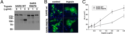
The coronavirus spike protein (S) plays a key role in the early steps of viral infection, with the S1 domain responsible for receptor binding and the S2 domain mediating membrane fusion. In some cases, the S protein is proteolytically cleaved at the S1-S2 boundary. In the case of the severe acute respiratory syndrome coronavirus (SARS-CoV), it has been shown that virus entry requires the endosomal protease cathepsin L; however, it was also found that infection of SARS-CoV could be strongly induced by trypsin treatment. Overall, in terms of how cleavage might activate membrane fusion, proteolytic processing of the SARS-CoV S protein remains unclear. Here, we identify a proteolytic cleavage site within the SARS-CoV S2 domain (S2', R797). Mutation of R797 specifically inhibited trypsin-dependent fusion in both cell-cell fusion and pseudovirion entry assays. We also introduced a furin cleavage site at both the S2' cleavage site within S2 793-KPTKR-797 (S2'), as well as at the junction of S1 and S2. Introduction of a furin cleavage site at the S2' position allowed trypsin-independent cell-cell fusion, which was strongly increased by the presence of a second furin cleavage site at the S1-S2 position. Taken together, these data suggest a novel priming mechanism for a viral fusion protein, with a critical proteolytic cleavage event on the SARS-CoV S protein at position 797 (S2'), acting in concert with the S1-S2 cleavage site to mediate membrane fusion and virus infectivity.


Conflict of interest statement

The authors declare no conflict of interest.

Figures

Fig. 1.
Fig. 1.
Role of the S1–S2 boundary in trypsin-mediated activation of SARS-CoV S membrane fusion. (A) BHK cells transiently expressing SARS-CoV S wild type (SARS wt) or the SARS-R667N mutant were treated with 1 μg/mL trypsin for 30 min or 1 h. Samples were analyzed by Western blot analysis, with the C-terminal part of the spike protein detected by using an anti-C9 antibody. (B) VeroE6 cells expressing SARS-CoV S wild type (SARS wt) or the SARS-R667N mutant were treated with 2 μg/mL trypsin for 20 min, then incubated for 40 min in absence of trypsin and analyzed by immunofluorescence microscopy using an anti-S monoclonal antibody (green), with nuclei counterstained with Hoechst 33548 (blue). (Magnification: 200×.) (C) BHK cells cotransfected with plasmids encoding the SARS-CoV S wild type (SARS wt) or SARS-R667N, as well as a plasmid encoding luciferase under the control of the T7 polymerase were overlaid with VeroE6 cells expressing the T7 polymerase. Cell–cell fusion was induced by treating the cells with increasing amount of trypsin for 20 min. At 5 h after fusion induction, the cells were lysed to monitor luciferase activity. Results are presented as relative light units (RLU). Error bars represent the standard error of the mean for 3 independent experiments.
Fig. 2.
Fig. 2.
Introduction of a furin site at S2′ induces SARS-CoV S-mediated membrane fusion. (A) VeroE6 cells expressing SARS-CoV S wild type (SARS wt) or the mutants SARS-Fur667, SARS-Fur797, or SARS-Fur667-797 were analyzed by immunofluorescence microscopy using an anti-S monoclonal antibody (green), with nuclei counterstained with Hoechst 33548 (blue). (Magnification: 200×.) (B) VeroE6 cells expressing SARS-CoV S wild type (SARS wt) or furin mutants were analyzed by immunofluorescence microscopy, and the number of nuclei involved in syncytia formation was counted for each mutant. Error bars represent the standard error of the mean for 3 independent experiments. Data were analyzed by using a Student's t test. (C) BHK cells were cotransfected with SARS-CoV S wild type (SARS wt) or furin mutants and with a T7-driven luciferase plasmid and then were overlaid with VeroE6 cells expressing the T7 polymerase. Luciferase activity was then measured after 8 h. Results are presented as relative light units (RLU). Error bars represent the standard error of the mean for 3 independent experiments. Data were analyzed by using a Student's t test. (D) BHK cells expressing SARS-CoV S wild type (SARS wt) or furin mutants were analyzed by Western blot analysis, with the C-terminal part of the spike protein detected by using an anti-myc antibody. (Lower) A longer exposure time of a portion of the Western blot analysis to more clearly show the cleavage products for the mutant furin797 (F797).
Fig. 3.
Fig. 3.
Mutation of R797 at SARS-CoV S2′ inhibits trypsin-induced membrane fusion. (A) VeroE6 cells expressing SARS-CoV S wild type (SARS wt) or the S2′ mutants were treated with 2 μg/mL trypsin for 20 min, then incubated for 40 min in the absence of trypsin and analyzed by immunofluorescence microscopy using an anti-S monoclonal antibody (green), with nuclei counterstained with Hoechst 33548 (blue). (Magnification: 200×.) (B) BHK cells, cotransfected with plasmids encoding the SARS-CoV S wild type (SARS wt) or mutants and a plasmid encoding luciferase under the control of the T7 polymerase, were overlaid with VeroE6 cells expressing the T7 polymerase. Cell–cell fusion was induced by treating the cells with 1 μg/mL trypsin for 20 min. At 5 h after fusion induction, the cells were lysed to monitor luciferase activity. Results are presented as relative light units (RLU). Error bars represent the standard error of the mean for 3 independent experiments. Data were analyzed by using an ANOVA test.
Fig. 4.
Fig. 4.
Role of S1/S2 and S2′ cleavage in virus entry mediated by SARS-CoV S. BHK cells coexpressing ACE2 and DC-SIGN were pretreated for 1 h with 25 mM NH4Cl. Virions pseudotyped with the SARS-CoV S wild type (SARS wt) or the different mutants were bound at 4 °C for 2 h in the presence of drug. Viral entry was induced by a 5-min treatment of the cells at 37 °C with trypsin. The results are expressed as the percentage of infection recovery obtained for the wild type. Error bars represent the standard error of the mean for 3 independent experiments. Data were analyzed by using a Student's t test.

References

    1. White JM, Delos SE, Brecher M, Schornberg K. Structures and mechanisms of viral membrane fusion proteins: multiple variations on a common theme. Crit Rev Biochem Mol Biol. 2008;43:189–219. - PMC - PubMed
    1. Harrison SC. Viral membrane fusion. Nat Struct Mol Biol. 2008;15:690–698. - PMC - PubMed
    1. Colman PM, Lawrence MC. The structural biology of type I viral membrane fusion. Nat Rev Mol Cell Biol. 2003;4:309–319. - PubMed
    1. Lamb RA, Jardetzky TS. Structural basis of viral invasion: Lessons from paramyxovirus F. Curr Opin Struct Biol. 2007;17:427–436. - PMC - PubMed
    1. Lai AL, Li Y, Tamm LK. In: Protein–Lipid Interactions. Tamm LK, editor. Weinheim, Germany: Wiley-VCH; 2005. pp. 279–303.

Publication types

MeSH terms

LinkOut - more resources